{"title":"罕见的卵巢黏液交界性肿瘤伴大实性成分。","authors":"Eito Kozawa, Kaiji Inoue, Mitsutake Yano, Masanori Yasuda, Kosei Hasegawa, Junji Tanaka, Tomoaki Ichikawa, Mamoru Niitsu","doi":"10.1155/2019/1402736","DOIUrl":null,"url":null,"abstract":"<p><p>Herein, we report magnetic resonance imaging (MRI) findings of a mucinous borderline tumor of the ovary, which we observed as a mainly solid tumor with large solid components in the lower pelvic cavity. The appearance of ovarian epithelial tumors on imaging is often complex. Cystic to solid appearing masses may be observed, and they often resemble epithelial carcinoma. Due to mucinous or hemorrhage components of packed small or microcystic components, MRI depicts slightly high signal intensity on T1-weighted images and low signal intensity on T2-weighted images. Mucinous borderline tumor of the ovary with a large solid component is very rare, but it is clinically important to recognize the possibility of mucinous borderline tumor to avoid unnecessary surgical intervention.</p>","PeriodicalId":30326,"journal":{"name":"Case Reports in Radiology","volume":" ","pages":"1402736"},"PeriodicalIF":0.0000,"publicationDate":"2019-05-22","publicationTypes":"Journal Article","fieldsOfStudy":null,"isOpenAccess":false,"openAccessPdf":"https://sci-hub-pdf.com/10.1155/2019/1402736","citationCount":"3","resultStr":"{\"title\":\"An Unusual Ovarian Mucinous Borderline Tumor with a Large Solid Component.\",\"authors\":\"Eito Kozawa, Kaiji Inoue, Mitsutake Yano, Masanori Yasuda, Kosei Hasegawa, Junji Tanaka, Tomoaki Ichikawa, Mamoru Niitsu\",\"doi\":\"10.1155/2019/1402736\",\"DOIUrl\":null,\"url\":null,\"abstract\":\"<p><p>Herein, we report magnetic resonance imaging (MRI) findings of a mucinous borderline tumor of the ovary, which we observed as a mainly solid tumor with large solid components in the lower pelvic cavity. The appearance of ovarian epithelial tumors on imaging is often complex. Cystic to solid appearing masses may be observed, and they often resemble epithelial carcinoma. Due to mucinous or hemorrhage components of packed small or microcystic components, MRI depicts slightly high signal intensity on T1-weighted images and low signal intensity on T2-weighted images. Mucinous borderline tumor of the ovary with a large solid component is very rare, but it is clinically important to recognize the possibility of mucinous borderline tumor to avoid unnecessary surgical intervention.</p>\",\"PeriodicalId\":30326,\"journal\":{\"name\":\"Case Reports in Radiology\",\"volume\":\" \",\"pages\":\"1402736\"},\"PeriodicalIF\":0.0000,\"publicationDate\":\"2019-05-22\",\"publicationTypes\":\"Journal Article\",\"fieldsOfStudy\":null,\"isOpenAccess\":false,\"openAccessPdf\":\"https://sci-hub-pdf.com/10.1155/2019/1402736\",\"citationCount\":\"3\",\"resultStr\":null,\"platform\":\"Semanticscholar\",\"paperid\":null,\"PeriodicalName\":\"Case Reports in Radiology\",\"FirstCategoryId\":\"1085\",\"ListUrlMain\":\"https://doi.org/10.1155/2019/1402736\",\"RegionNum\":0,\"RegionCategory\":null,\"ArticlePicture\":[],\"TitleCN\":null,\"AbstractTextCN\":null,\"PMCID\":null,\"EPubDate\":\"2019/1/1 0:00:00\",\"PubModel\":\"eCollection\",\"JCR\":\"\",\"JCRName\":\"\",\"Score\":null,\"Total\":0}","platform":"Semanticscholar","paperid":null,"PeriodicalName":"Case Reports in Radiology","FirstCategoryId":"1085","ListUrlMain":"https://doi.org/10.1155/2019/1402736","RegionNum":0,"RegionCategory":null,"ArticlePicture":[],"TitleCN":null,"AbstractTextCN":null,"PMCID":null,"EPubDate":"2019/1/1 0:00:00","PubModel":"eCollection","JCR":"","JCRName":"","Score":null,"Total":0}
An Unusual Ovarian Mucinous Borderline Tumor with a Large Solid Component.
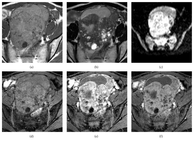
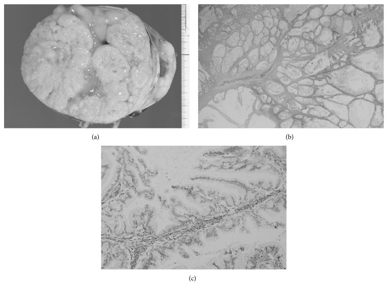
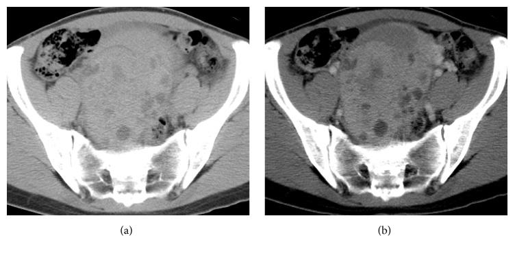
Herein, we report magnetic resonance imaging (MRI) findings of a mucinous borderline tumor of the ovary, which we observed as a mainly solid tumor with large solid components in the lower pelvic cavity. The appearance of ovarian epithelial tumors on imaging is often complex. Cystic to solid appearing masses may be observed, and they often resemble epithelial carcinoma. Due to mucinous or hemorrhage components of packed small or microcystic components, MRI depicts slightly high signal intensity on T1-weighted images and low signal intensity on T2-weighted images. Mucinous borderline tumor of the ovary with a large solid component is very rare, but it is clinically important to recognize the possibility of mucinous borderline tumor to avoid unnecessary surgical intervention.

 求助内容:
求助内容: 应助结果提醒方式:


