{"title":"老年痴呆症的老年科学小鼠模型。","authors":"Martin Darvas, Dirk Keene, Warren Ladiges","doi":"10.1080/20010001.2019.1616994","DOIUrl":null,"url":null,"abstract":"Geroscience is a multidisciplinary field that examines the relationship between biological aging and agerelated diseases [1]. Seven processes discussed by the trans-NIH Geroscience Interest Group Summit that contribute to biological aging included macromolecular damage, epigenetic changes, inflammation, adaptation to stress, impairments to proteostasis, stem cell regeneration, and metabolism [2]. These processes are highly integrated with one another such that targeting them as a group may be an effective approach to developing therapies to prevent or delay age-related disease. Alzheimer’s disease (AD) is an age-related disease and is expected to increase with the number of elderly individuals rapidly rising in both developed and developing countries. Efforts to find diseasemodifying treatments have met with limited success possibly because they have focused on identifying a specific pathogenic mechanism targeted by a specific drug. AD is a complex disease involving numerous mechanisms in line with processes of biological aging. Therefore, a geroscience approach to successfully treating AD is a logical concept that unfortunately has not yet been widely accepted by the neuroscience community. It is now time to explore preclinical studies in AD animal models to begin screening different drug combinations that target multiple aging-related processes for effect on AD dementia and neuropathology. A major challenge for preclinical drug testing is the selection of an AD animal model. A model is needed that shows amyloid (A) β and tau neuropathology, inflammation, oxidative stress, neuronal degeneration, and neurovascular deficits in an aging background. Currently available models are transgenic mice expressing amyloid precursor protein (APP) and presenilin mutations found in patients with early onset of AD. These mouse models are useful, but develop lesions at an early age, and none represent all the mechanisms representative of human AD. Ideally, the model should be easily manipulated so that dementia and neuropathology can be induced in an old-age animal as well as middle age, and young age to compare disease progression in different aging backgrounds. The animal of choice for large-scale drug testing is the mouse, but the rat could also be considered. There are advantages and disadvantages for both but our lab has extensive experience with aging mice so the aging mouse will be the prototype animal for this discussion. Aging in mice is in many ways similar to aging in people, so the geroscience concept is applicable, as depicted in Figure 1. The two hall-mark molecular pathologic changes of AD are accumulation of amyloid β 42 peptides (Aβ42) and paired helical filament (PHF)-tau (τ). Aβ42 pathology is not evenly distributed, but systematically localized to certain parts of the brain, following a prototypical sequence in which the regions are hierarchically involved: cortical Aβ42 deposits, followed by involvement of allocortical regions, involvement of subcortical forebrain regions and striatum, deposits in the brainstem and finally cerebellar Aβ42-deposition. The distribution of Aβ42 across the brain is part of the current NIA-AA consensus criteria for the neuropathologic diagnosis of AD [3]. To allow control over the extent of Aβ42 expression and better alignment with the clinical distribution of Aβ42, an ADmodel that is based on adeno-associated virus (AAV) mediated Aβ42 expression was developed and validated in adult rats [4]. This model overcomes the shortcomings of current transgenic models of AD pathology because it allows induction of pathology at a disease-relevant age and restricts pathology to injected brain regions selected based on their relevance to AD. While genetic data clearly support mutations in the APP gene as sufficient to produce AD, and extensive experimental evidence points to the neurotoxicity of Aβ peptides, it is now appreciated that cerebral cortical accumulation of fibrillar Aβ occurs in virtually all older adults and reaches maximal concentration in the preclinical or prodromal stages of AD prior to clinical expression of dementia. In contrast, AD dementia is closely associated with the extension of PHF-τ beyond mesial temporal lobe structures. The distribution of PHF-τ pathology in AD also follows a prototypical sequence with hierarchical involvement of the following regions: transentorhinal cortical layer, entorhinal layer and hippocampus, and isocortical PHF-τ containing tangles. Similar to Aβ42, the distribution pattern of PHF-τ is part of the current NIA-AA consensus criteria for the neuropathologic diagnosis of AD [3]. Therefore, we also include the formation of PHF-τ in AAV-based models of AD. Although there are no known associations between mutations in the gene encoding τ (MAPT) and AD, there are strong associations between mutations in MAPT and hereditary frontotemporal PATHOBIOLOGY OF AGING & AGE-RELATED DISEASES 2019, VOL. 9, 1616994 https://doi.org/10.1080/20010001.2019.1616994","PeriodicalId":89611,"journal":{"name":"Pathobiology of aging & age related diseases","volume":"9 1","pages":"1616994"},"PeriodicalIF":0.0000,"publicationDate":"2019-05-14","publicationTypes":"Journal Article","fieldsOfStudy":null,"isOpenAccess":false,"openAccessPdf":"https://sci-hub-pdf.com/10.1080/20010001.2019.1616994","citationCount":"3","resultStr":"{\"title\":\"A geroscience mouse model for Alzheimer's disease.\",\"authors\":\"Martin Darvas, Dirk Keene, Warren Ladiges\",\"doi\":\"10.1080/20010001.2019.1616994\",\"DOIUrl\":null,\"url\":null,\"abstract\":\"Geroscience is a multidisciplinary field that examines the relationship between biological aging and agerelated diseases [1]. Seven processes discussed by the trans-NIH Geroscience Interest Group Summit that contribute to biological aging included macromolecular damage, epigenetic changes, inflammation, adaptation to stress, impairments to proteostasis, stem cell regeneration, and metabolism [2]. These processes are highly integrated with one another such that targeting them as a group may be an effective approach to developing therapies to prevent or delay age-related disease. Alzheimer’s disease (AD) is an age-related disease and is expected to increase with the number of elderly individuals rapidly rising in both developed and developing countries. Efforts to find diseasemodifying treatments have met with limited success possibly because they have focused on identifying a specific pathogenic mechanism targeted by a specific drug. AD is a complex disease involving numerous mechanisms in line with processes of biological aging. Therefore, a geroscience approach to successfully treating AD is a logical concept that unfortunately has not yet been widely accepted by the neuroscience community. It is now time to explore preclinical studies in AD animal models to begin screening different drug combinations that target multiple aging-related processes for effect on AD dementia and neuropathology. A major challenge for preclinical drug testing is the selection of an AD animal model. A model is needed that shows amyloid (A) β and tau neuropathology, inflammation, oxidative stress, neuronal degeneration, and neurovascular deficits in an aging background. Currently available models are transgenic mice expressing amyloid precursor protein (APP) and presenilin mutations found in patients with early onset of AD. These mouse models are useful, but develop lesions at an early age, and none represent all the mechanisms representative of human AD. Ideally, the model should be easily manipulated so that dementia and neuropathology can be induced in an old-age animal as well as middle age, and young age to compare disease progression in different aging backgrounds. The animal of choice for large-scale drug testing is the mouse, but the rat could also be considered. There are advantages and disadvantages for both but our lab has extensive experience with aging mice so the aging mouse will be the prototype animal for this discussion. Aging in mice is in many ways similar to aging in people, so the geroscience concept is applicable, as depicted in Figure 1. The two hall-mark molecular pathologic changes of AD are accumulation of amyloid β 42 peptides (Aβ42) and paired helical filament (PHF)-tau (τ). Aβ42 pathology is not evenly distributed, but systematically localized to certain parts of the brain, following a prototypical sequence in which the regions are hierarchically involved: cortical Aβ42 deposits, followed by involvement of allocortical regions, involvement of subcortical forebrain regions and striatum, deposits in the brainstem and finally cerebellar Aβ42-deposition. The distribution of Aβ42 across the brain is part of the current NIA-AA consensus criteria for the neuropathologic diagnosis of AD [3]. To allow control over the extent of Aβ42 expression and better alignment with the clinical distribution of Aβ42, an ADmodel that is based on adeno-associated virus (AAV) mediated Aβ42 expression was developed and validated in adult rats [4]. This model overcomes the shortcomings of current transgenic models of AD pathology because it allows induction of pathology at a disease-relevant age and restricts pathology to injected brain regions selected based on their relevance to AD. While genetic data clearly support mutations in the APP gene as sufficient to produce AD, and extensive experimental evidence points to the neurotoxicity of Aβ peptides, it is now appreciated that cerebral cortical accumulation of fibrillar Aβ occurs in virtually all older adults and reaches maximal concentration in the preclinical or prodromal stages of AD prior to clinical expression of dementia. In contrast, AD dementia is closely associated with the extension of PHF-τ beyond mesial temporal lobe structures. The distribution of PHF-τ pathology in AD also follows a prototypical sequence with hierarchical involvement of the following regions: transentorhinal cortical layer, entorhinal layer and hippocampus, and isocortical PHF-τ containing tangles. Similar to Aβ42, the distribution pattern of PHF-τ is part of the current NIA-AA consensus criteria for the neuropathologic diagnosis of AD [3]. Therefore, we also include the formation of PHF-τ in AAV-based models of AD. Although there are no known associations between mutations in the gene encoding τ (MAPT) and AD, there are strong associations between mutations in MAPT and hereditary frontotemporal PATHOBIOLOGY OF AGING & AGE-RELATED DISEASES 2019, VOL. 9, 1616994 https://doi.org/10.1080/20010001.2019.1616994\",\"PeriodicalId\":89611,\"journal\":{\"name\":\"Pathobiology of aging & age related diseases\",\"volume\":\"9 1\",\"pages\":\"1616994\"},\"PeriodicalIF\":0.0000,\"publicationDate\":\"2019-05-14\",\"publicationTypes\":\"Journal Article\",\"fieldsOfStudy\":null,\"isOpenAccess\":false,\"openAccessPdf\":\"https://sci-hub-pdf.com/10.1080/20010001.2019.1616994\",\"citationCount\":\"3\",\"resultStr\":null,\"platform\":\"Semanticscholar\",\"paperid\":null,\"PeriodicalName\":\"Pathobiology of aging & age related diseases\",\"FirstCategoryId\":\"1085\",\"ListUrlMain\":\"https://doi.org/10.1080/20010001.2019.1616994\",\"RegionNum\":0,\"RegionCategory\":null,\"ArticlePicture\":[],\"TitleCN\":null,\"AbstractTextCN\":null,\"PMCID\":null,\"EPubDate\":\"2019/1/1 0:00:00\",\"PubModel\":\"eCollection\",\"JCR\":\"\",\"JCRName\":\"\",\"Score\":null,\"Total\":0}","platform":"Semanticscholar","paperid":null,"PeriodicalName":"Pathobiology of aging & age related diseases","FirstCategoryId":"1085","ListUrlMain":"https://doi.org/10.1080/20010001.2019.1616994","RegionNum":0,"RegionCategory":null,"ArticlePicture":[],"TitleCN":null,"AbstractTextCN":null,"PMCID":null,"EPubDate":"2019/1/1 0:00:00","PubModel":"eCollection","JCR":"","JCRName":"","Score":null,"Total":0}
A geroscience mouse model for Alzheimer's disease.
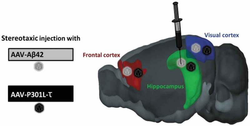
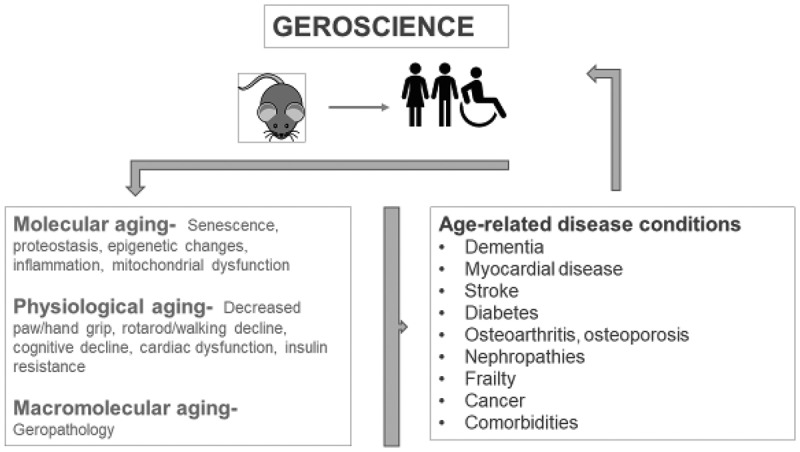
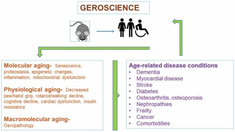
Geroscience is a multidisciplinary field that examines the relationship between biological aging and agerelated diseases [1]. Seven processes discussed by the trans-NIH Geroscience Interest Group Summit that contribute to biological aging included macromolecular damage, epigenetic changes, inflammation, adaptation to stress, impairments to proteostasis, stem cell regeneration, and metabolism [2]. These processes are highly integrated with one another such that targeting them as a group may be an effective approach to developing therapies to prevent or delay age-related disease. Alzheimer’s disease (AD) is an age-related disease and is expected to increase with the number of elderly individuals rapidly rising in both developed and developing countries. Efforts to find diseasemodifying treatments have met with limited success possibly because they have focused on identifying a specific pathogenic mechanism targeted by a specific drug. AD is a complex disease involving numerous mechanisms in line with processes of biological aging. Therefore, a geroscience approach to successfully treating AD is a logical concept that unfortunately has not yet been widely accepted by the neuroscience community. It is now time to explore preclinical studies in AD animal models to begin screening different drug combinations that target multiple aging-related processes for effect on AD dementia and neuropathology. A major challenge for preclinical drug testing is the selection of an AD animal model. A model is needed that shows amyloid (A) β and tau neuropathology, inflammation, oxidative stress, neuronal degeneration, and neurovascular deficits in an aging background. Currently available models are transgenic mice expressing amyloid precursor protein (APP) and presenilin mutations found in patients with early onset of AD. These mouse models are useful, but develop lesions at an early age, and none represent all the mechanisms representative of human AD. Ideally, the model should be easily manipulated so that dementia and neuropathology can be induced in an old-age animal as well as middle age, and young age to compare disease progression in different aging backgrounds. The animal of choice for large-scale drug testing is the mouse, but the rat could also be considered. There are advantages and disadvantages for both but our lab has extensive experience with aging mice so the aging mouse will be the prototype animal for this discussion. Aging in mice is in many ways similar to aging in people, so the geroscience concept is applicable, as depicted in Figure 1. The two hall-mark molecular pathologic changes of AD are accumulation of amyloid β 42 peptides (Aβ42) and paired helical filament (PHF)-tau (τ). Aβ42 pathology is not evenly distributed, but systematically localized to certain parts of the brain, following a prototypical sequence in which the regions are hierarchically involved: cortical Aβ42 deposits, followed by involvement of allocortical regions, involvement of subcortical forebrain regions and striatum, deposits in the brainstem and finally cerebellar Aβ42-deposition. The distribution of Aβ42 across the brain is part of the current NIA-AA consensus criteria for the neuropathologic diagnosis of AD [3]. To allow control over the extent of Aβ42 expression and better alignment with the clinical distribution of Aβ42, an ADmodel that is based on adeno-associated virus (AAV) mediated Aβ42 expression was developed and validated in adult rats [4]. This model overcomes the shortcomings of current transgenic models of AD pathology because it allows induction of pathology at a disease-relevant age and restricts pathology to injected brain regions selected based on their relevance to AD. While genetic data clearly support mutations in the APP gene as sufficient to produce AD, and extensive experimental evidence points to the neurotoxicity of Aβ peptides, it is now appreciated that cerebral cortical accumulation of fibrillar Aβ occurs in virtually all older adults and reaches maximal concentration in the preclinical or prodromal stages of AD prior to clinical expression of dementia. In contrast, AD dementia is closely associated with the extension of PHF-τ beyond mesial temporal lobe structures. The distribution of PHF-τ pathology in AD also follows a prototypical sequence with hierarchical involvement of the following regions: transentorhinal cortical layer, entorhinal layer and hippocampus, and isocortical PHF-τ containing tangles. Similar to Aβ42, the distribution pattern of PHF-τ is part of the current NIA-AA consensus criteria for the neuropathologic diagnosis of AD [3]. Therefore, we also include the formation of PHF-τ in AAV-based models of AD. Although there are no known associations between mutations in the gene encoding τ (MAPT) and AD, there are strong associations between mutations in MAPT and hereditary frontotemporal PATHOBIOLOGY OF AGING & AGE-RELATED DISEASES 2019, VOL. 9, 1616994 https://doi.org/10.1080/20010001.2019.1616994

 求助内容:
求助内容: 应助结果提醒方式:


