Mohammad Ali Ansari, Mohsen Mohebati, Farid Poursadegh, Mahdi Foroughian, Alireza Sepehri Shamloo
{"title":"超声心动图显示冠心病患者心外膜脂肪厚度增加吗?系统回顾和荟萃分析。","authors":"Mohammad Ali Ansari, Mohsen Mohebati, Farid Poursadegh, Mahdi Foroughian, Alireza Sepehri Shamloo","doi":"10.19082/7249","DOIUrl":null,"url":null,"abstract":"<p><strong>Background: </strong>The relation of epicardial fat thickness (EFT) to coronary artery disease (CAD) has recently been reported in multiple studies. Echocardiography is a safe and relatively inexpensive and accessible approach to assess regional EFT, which can be performed easily in many centers.</p><p><strong>Objective: </strong>To determine the association between echocardiographic EFT and the presence or the absence of CAD.</p><p><strong>Methods: </strong>This was a systematic review and meta-analysis conducted on literature available in electronic databases up to March 2018. The articles measuring EFT by echocardiography in the right ventricular (RV) free wall were included in the study. The quality of the enrolled items was assessed using the Methodological Index for Non-Randomized Studies (MINORS) checklist. The analyses were performed using the Comprehensive Meta-Analysis version 2 software. Cochran's Q test and I<sup>2</sup> index were used to evaluate heterogeneity.</p><p><strong>Results: </strong>This meta-analysis was performed on 13 studies involving 2,436 patients (1,622 with CAD, and 814 without CAD). The maximum EFT reported by echocardiography was 12.9±2.7 mm in the CAD group and 8.4±2.5 mm in the non-CAD group. The minimum EFT reported by echocardiography was 2.2±1.8 mm in the CAD group and 1.8±1.4 mm in the non-CAD group. The heterogeneity was found among the researched studies (I<sup>2</sup>=91.8%, p=0.000, Q-value=146.43, df [Q] =12) using the random effect model. The patients with CAD had a significantly higher echocardiographic EFT than those without CAD (SMD=1.03, 95% CI= 0.70-1.37, p=0.000).</p><p><strong>Conclusion: </strong>According to the findings of this meta-analysis, the echocardiographic EFT in the subjects with CAD was significantly higher than that of those without CAD. The measurement of echocardiographic EFT seems to be an acceptable strategy for risk stratification of heart diseases considering ease of use, cost-effectiveness and non-exposure characteristics, compared to other imaging interventions.</p>","PeriodicalId":11603,"journal":{"name":"Electronic Physician","volume":"10 9","pages":"7249-7258"},"PeriodicalIF":0.0000,"publicationDate":"2018-09-09","publicationTypes":"Journal Article","fieldsOfStudy":null,"isOpenAccess":false,"openAccessPdf":"https://ftp.ncbi.nlm.nih.gov/pub/pmc/oa_pdf/f9/d7/epj-10-7249.PMC6140987.pdf","citationCount":"13","resultStr":"{\"title\":\"Is echocardiographic epicardial fat thickness increased in patients with coronary artery disease? A systematic review and meta-analysis.\",\"authors\":\"Mohammad Ali Ansari, Mohsen Mohebati, Farid Poursadegh, Mahdi Foroughian, Alireza Sepehri Shamloo\",\"doi\":\"10.19082/7249\",\"DOIUrl\":null,\"url\":null,\"abstract\":\"<p><strong>Background: </strong>The relation of epicardial fat thickness (EFT) to coronary artery disease (CAD) has recently been reported in multiple studies. Echocardiography is a safe and relatively inexpensive and accessible approach to assess regional EFT, which can be performed easily in many centers.</p><p><strong>Objective: </strong>To determine the association between echocardiographic EFT and the presence or the absence of CAD.</p><p><strong>Methods: </strong>This was a systematic review and meta-analysis conducted on literature available in electronic databases up to March 2018. The articles measuring EFT by echocardiography in the right ventricular (RV) free wall were included in the study. The quality of the enrolled items was assessed using the Methodological Index for Non-Randomized Studies (MINORS) checklist. The analyses were performed using the Comprehensive Meta-Analysis version 2 software. Cochran's Q test and I<sup>2</sup> index were used to evaluate heterogeneity.</p><p><strong>Results: </strong>This meta-analysis was performed on 13 studies involving 2,436 patients (1,622 with CAD, and 814 without CAD). The maximum EFT reported by echocardiography was 12.9±2.7 mm in the CAD group and 8.4±2.5 mm in the non-CAD group. The minimum EFT reported by echocardiography was 2.2±1.8 mm in the CAD group and 1.8±1.4 mm in the non-CAD group. The heterogeneity was found among the researched studies (I<sup>2</sup>=91.8%, p=0.000, Q-value=146.43, df [Q] =12) using the random effect model. The patients with CAD had a significantly higher echocardiographic EFT than those without CAD (SMD=1.03, 95% CI= 0.70-1.37, p=0.000).</p><p><strong>Conclusion: </strong>According to the findings of this meta-analysis, the echocardiographic EFT in the subjects with CAD was significantly higher than that of those without CAD. The measurement of echocardiographic EFT seems to be an acceptable strategy for risk stratification of heart diseases considering ease of use, cost-effectiveness and non-exposure characteristics, compared to other imaging interventions.</p>\",\"PeriodicalId\":11603,\"journal\":{\"name\":\"Electronic Physician\",\"volume\":\"10 9\",\"pages\":\"7249-7258\"},\"PeriodicalIF\":0.0000,\"publicationDate\":\"2018-09-09\",\"publicationTypes\":\"Journal Article\",\"fieldsOfStudy\":null,\"isOpenAccess\":false,\"openAccessPdf\":\"https://ftp.ncbi.nlm.nih.gov/pub/pmc/oa_pdf/f9/d7/epj-10-7249.PMC6140987.pdf\",\"citationCount\":\"13\",\"resultStr\":null,\"platform\":\"Semanticscholar\",\"paperid\":null,\"PeriodicalName\":\"Electronic Physician\",\"FirstCategoryId\":\"1085\",\"ListUrlMain\":\"https://doi.org/10.19082/7249\",\"RegionNum\":0,\"RegionCategory\":null,\"ArticlePicture\":[],\"TitleCN\":null,\"AbstractTextCN\":null,\"PMCID\":null,\"EPubDate\":\"2018/9/1 0:00:00\",\"PubModel\":\"eCollection\",\"JCR\":\"\",\"JCRName\":\"\",\"Score\":null,\"Total\":0}","platform":"Semanticscholar","paperid":null,"PeriodicalName":"Electronic Physician","FirstCategoryId":"1085","ListUrlMain":"https://doi.org/10.19082/7249","RegionNum":0,"RegionCategory":null,"ArticlePicture":[],"TitleCN":null,"AbstractTextCN":null,"PMCID":null,"EPubDate":"2018/9/1 0:00:00","PubModel":"eCollection","JCR":"","JCRName":"","Score":null,"Total":0}
Is echocardiographic epicardial fat thickness increased in patients with coronary artery disease? A systematic review and meta-analysis.
Background: The relation of epicardial fat thickness (EFT) to coronary artery disease (CAD) has recently been reported in multiple studies. Echocardiography is a safe and relatively inexpensive and accessible approach to assess regional EFT, which can be performed easily in many centers.
Objective: To determine the association between echocardiographic EFT and the presence or the absence of CAD.
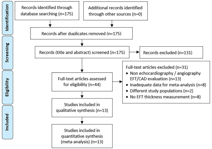
Methods: This was a systematic review and meta-analysis conducted on literature available in electronic databases up to March 2018. The articles measuring EFT by echocardiography in the right ventricular (RV) free wall were included in the study. The quality of the enrolled items was assessed using the Methodological Index for Non-Randomized Studies (MINORS) checklist. The analyses were performed using the Comprehensive Meta-Analysis version 2 software. Cochran's Q test and I2 index were used to evaluate heterogeneity.
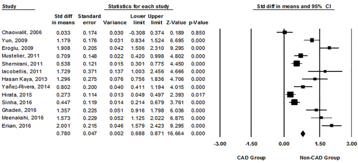
Results: This meta-analysis was performed on 13 studies involving 2,436 patients (1,622 with CAD, and 814 without CAD). The maximum EFT reported by echocardiography was 12.9±2.7 mm in the CAD group and 8.4±2.5 mm in the non-CAD group. The minimum EFT reported by echocardiography was 2.2±1.8 mm in the CAD group and 1.8±1.4 mm in the non-CAD group. The heterogeneity was found among the researched studies (I2=91.8%, p=0.000, Q-value=146.43, df [Q] =12) using the random effect model. The patients with CAD had a significantly higher echocardiographic EFT than those without CAD (SMD=1.03, 95% CI= 0.70-1.37, p=0.000).
Conclusion: According to the findings of this meta-analysis, the echocardiographic EFT in the subjects with CAD was significantly higher than that of those without CAD. The measurement of echocardiographic EFT seems to be an acceptable strategy for risk stratification of heart diseases considering ease of use, cost-effectiveness and non-exposure characteristics, compared to other imaging interventions.

 求助内容:
求助内容: 应助结果提醒方式:


