Lei Zhang, Yawei Zhang, You Zhang, Wendy B Harris, Fang-Fang Yin, Jing Cai, Lei Ren
{"title":"基于先验知识和患者运动建模的无标记四维锥束计算机断层扫描投影相位排序的可行性研究。","authors":"Lei Zhang, Yawei Zhang, You Zhang, Wendy B Harris, Fang-Fang Yin, Jing Cai, Lei Ren","doi":"","DOIUrl":null,"url":null,"abstract":"<p><strong>Aim: </strong>During cancer radiotherapy treatment, on-board four-dimensional-cone beam computed tomography (4D-CBCT) provides important patient 4D volumetric information for tumor target verification. Reconstruction of 4D-CBCT images requires sorting of acquired projections into different respiratory phases. Traditional phase sorting methods are either based on external surrogates, which might miscorrelate with internal structures; or on 2D internal structures, which require specific organ presence or slow gantry rotations. The aim of this study is to investigate the feasibility of a 3D motion modeling-based method for markerless 4D-CBCT projection-phase sorting.</p><p><strong>Methods: </strong>Patient 4D-CT images acquired during simulation are used as prior images. Principal component analysis (PCA) is used to extract three major respiratory deformation patterns. On-board patient image volume is considered as a deformation of the prior CT at the end-expiration phase. Coefficients of the principal deformation patterns are solved for each on-board projection by matching it with the digitally reconstructed radiograph (DRR) of the deformed prior CT. The primary PCA coefficients are used for the projection-phase sorting.</p><p><strong>Results: </strong>PCA coefficients solved in nine digital phantoms (XCATs) showed the same pattern as the breathing motions in both the anteroposterior and superoinferior directions. The mean phase sorting differences were below 2% and percentages of phase difference < 10% were 100% for all the nine XCAT phantoms. Five lung cancer patient results showed mean phase difference ranging from 1.62% to 2.23%. The percentage of projections within 10% phase difference ranged from 98.4% to 100% and those within 5% phase difference ranged from 88.9% to 99.8%.</p><p><strong>Conclusion: </strong>The study demonstrated the feasibility of using PCA coefficients for 4D-CBCT projection-phase sorting. High sorting accuracy in both digital phantoms and patient cases was achieved. This method provides an accurate and robust tool for automatic 4D-CBCT projection sorting using 3D motion modeling without the need of external surrogate or internal markers.</p>","PeriodicalId":9428,"journal":{"name":"Cancer Translational Medicine","volume":"3 6","pages":"185-193"},"PeriodicalIF":0.0000,"publicationDate":"2017-01-01","publicationTypes":"Journal Article","fieldsOfStudy":null,"isOpenAccess":false,"openAccessPdf":"https://ftp.ncbi.nlm.nih.gov/pub/pmc/oa_pdf/1b/80/nihms945805.PMC6101251.pdf","citationCount":"0","resultStr":"{\"title\":\"Markerless Four-Dimensional-Cone Beam Computed Tomography Projection-Phase Sorting Using Prior Knowledge and Patient Motion Modeling: A Feasibility Study.\",\"authors\":\"Lei Zhang, Yawei Zhang, You Zhang, Wendy B Harris, Fang-Fang Yin, Jing Cai, Lei Ren\",\"doi\":\"\",\"DOIUrl\":null,\"url\":null,\"abstract\":\"<p><strong>Aim: </strong>During cancer radiotherapy treatment, on-board four-dimensional-cone beam computed tomography (4D-CBCT) provides important patient 4D volumetric information for tumor target verification. Reconstruction of 4D-CBCT images requires sorting of acquired projections into different respiratory phases. Traditional phase sorting methods are either based on external surrogates, which might miscorrelate with internal structures; or on 2D internal structures, which require specific organ presence or slow gantry rotations. The aim of this study is to investigate the feasibility of a 3D motion modeling-based method for markerless 4D-CBCT projection-phase sorting.</p><p><strong>Methods: </strong>Patient 4D-CT images acquired during simulation are used as prior images. Principal component analysis (PCA) is used to extract three major respiratory deformation patterns. On-board patient image volume is considered as a deformation of the prior CT at the end-expiration phase. Coefficients of the principal deformation patterns are solved for each on-board projection by matching it with the digitally reconstructed radiograph (DRR) of the deformed prior CT. The primary PCA coefficients are used for the projection-phase sorting.</p><p><strong>Results: </strong>PCA coefficients solved in nine digital phantoms (XCATs) showed the same pattern as the breathing motions in both the anteroposterior and superoinferior directions. The mean phase sorting differences were below 2% and percentages of phase difference < 10% were 100% for all the nine XCAT phantoms. Five lung cancer patient results showed mean phase difference ranging from 1.62% to 2.23%. The percentage of projections within 10% phase difference ranged from 98.4% to 100% and those within 5% phase difference ranged from 88.9% to 99.8%.</p><p><strong>Conclusion: </strong>The study demonstrated the feasibility of using PCA coefficients for 4D-CBCT projection-phase sorting. High sorting accuracy in both digital phantoms and patient cases was achieved. This method provides an accurate and robust tool for automatic 4D-CBCT projection sorting using 3D motion modeling without the need of external surrogate or internal markers.</p>\",\"PeriodicalId\":9428,\"journal\":{\"name\":\"Cancer Translational Medicine\",\"volume\":\"3 6\",\"pages\":\"185-193\"},\"PeriodicalIF\":0.0000,\"publicationDate\":\"2017-01-01\",\"publicationTypes\":\"Journal Article\",\"fieldsOfStudy\":null,\"isOpenAccess\":false,\"openAccessPdf\":\"https://ftp.ncbi.nlm.nih.gov/pub/pmc/oa_pdf/1b/80/nihms945805.PMC6101251.pdf\",\"citationCount\":\"0\",\"resultStr\":null,\"platform\":\"Semanticscholar\",\"paperid\":null,\"PeriodicalName\":\"Cancer Translational Medicine\",\"FirstCategoryId\":\"1085\",\"ListUrlMain\":\"\",\"RegionNum\":0,\"RegionCategory\":null,\"ArticlePicture\":[],\"TitleCN\":null,\"AbstractTextCN\":null,\"PMCID\":null,\"EPubDate\":\"2017/12/29 0:00:00\",\"PubModel\":\"Epub\",\"JCR\":\"\",\"JCRName\":\"\",\"Score\":null,\"Total\":0}","platform":"Semanticscholar","paperid":null,"PeriodicalName":"Cancer Translational Medicine","FirstCategoryId":"1085","ListUrlMain":"","RegionNum":0,"RegionCategory":null,"ArticlePicture":[],"TitleCN":null,"AbstractTextCN":null,"PMCID":null,"EPubDate":"2017/12/29 0:00:00","PubModel":"Epub","JCR":"","JCRName":"","Score":null,"Total":0}
Markerless Four-Dimensional-Cone Beam Computed Tomography Projection-Phase Sorting Using Prior Knowledge and Patient Motion Modeling: A Feasibility Study.
Aim: During cancer radiotherapy treatment, on-board four-dimensional-cone beam computed tomography (4D-CBCT) provides important patient 4D volumetric information for tumor target verification. Reconstruction of 4D-CBCT images requires sorting of acquired projections into different respiratory phases. Traditional phase sorting methods are either based on external surrogates, which might miscorrelate with internal structures; or on 2D internal structures, which require specific organ presence or slow gantry rotations. The aim of this study is to investigate the feasibility of a 3D motion modeling-based method for markerless 4D-CBCT projection-phase sorting.
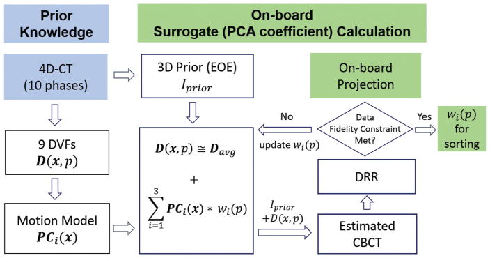
Methods: Patient 4D-CT images acquired during simulation are used as prior images. Principal component analysis (PCA) is used to extract three major respiratory deformation patterns. On-board patient image volume is considered as a deformation of the prior CT at the end-expiration phase. Coefficients of the principal deformation patterns are solved for each on-board projection by matching it with the digitally reconstructed radiograph (DRR) of the deformed prior CT. The primary PCA coefficients are used for the projection-phase sorting.
Results: PCA coefficients solved in nine digital phantoms (XCATs) showed the same pattern as the breathing motions in both the anteroposterior and superoinferior directions. The mean phase sorting differences were below 2% and percentages of phase difference < 10% were 100% for all the nine XCAT phantoms. Five lung cancer patient results showed mean phase difference ranging from 1.62% to 2.23%. The percentage of projections within 10% phase difference ranged from 98.4% to 100% and those within 5% phase difference ranged from 88.9% to 99.8%.
Conclusion: The study demonstrated the feasibility of using PCA coefficients for 4D-CBCT projection-phase sorting. High sorting accuracy in both digital phantoms and patient cases was achieved. This method provides an accurate and robust tool for automatic 4D-CBCT projection sorting using 3D motion modeling without the need of external surrogate or internal markers.


 求助内容:
求助内容: 应助结果提醒方式:


