Lauro Augusto Veloso Costa , Marcos Barbieri Mestriner , Thiago Alvim do Amaral , Bárbara dos Santos Barbosa , Camila Cohen Kaleka , Ricardo de Paula Leite Cury
{"title":"股后交叉韧带止点的组织学研究","authors":"Lauro Augusto Veloso Costa , Marcos Barbieri Mestriner , Thiago Alvim do Amaral , Bárbara dos Santos Barbosa , Camila Cohen Kaleka , Ricardo de Paula Leite Cury","doi":"10.1016/j.rboe.2018.05.006","DOIUrl":null,"url":null,"abstract":"<div><h3>Objectives</h3><p>To describe the microscopic anatomy of the posterior cruciate ligament femoral insertion in order to identify and establish differences between the direct and indirect insertions of this ligament.</p></div><div><h3>Methods</h3><p>Ten cadaveric knees were used for this study. The posterior cruciate ligament femoral insertion was observed microscopically. Hematoxylin and eosin staining was performed to observe the morphology of the posterior cruciate ligament insertion. Alcian blue staining was performed to determine the location of the cartilage matrix and better assist in the observation and differentiation between direct and indirect insertions.</p></div><div><h3>Results</h3><p>The direct insertion was observed to be a more complex structure than the indirect insertion because it showed four different histological layers (ligament, uncalcified fibrocartilage, calcified fibrocartilage, and bone). Chondrocytes were observed in the uncalcified and calcified fibrocartilage layers. It was observed that the indirect insertion was composed of two layers in which the ligament was anchored directly to the bone by collagen fibers. Indirect insertion was located in the marginal region of the posterior cruciate ligament between the direct insertion and the anterior articular cartilage.</p></div><div><h3>Conclusion</h3><p>Through histological analysis, it was demonstrated that the indirect insertion was adjacent to the anterior articular cartilage and presents a histological pattern where the collagen fibers insert directly into the bone (two-layer insertion). The direct insertion is posterior to the indirect insertion and has four histologically distinct layers.</p></div>","PeriodicalId":101095,"journal":{"name":"Revista Brasileira de Ortopedia (English Edition)","volume":"53 4","pages":"Pages 415-420"},"PeriodicalIF":0.0000,"publicationDate":"2018-07-01","publicationTypes":"Journal Article","fieldsOfStudy":null,"isOpenAccess":false,"openAccessPdf":"https://sci-hub-pdf.com/10.1016/j.rboe.2018.05.006","citationCount":"4","resultStr":"{\"title\":\"Histological study of the posterior cruciate ligament femoral insertion\",\"authors\":\"Lauro Augusto Veloso Costa , Marcos Barbieri Mestriner , Thiago Alvim do Amaral , Bárbara dos Santos Barbosa , Camila Cohen Kaleka , Ricardo de Paula Leite Cury\",\"doi\":\"10.1016/j.rboe.2018.05.006\",\"DOIUrl\":null,\"url\":null,\"abstract\":\"<div><h3>Objectives</h3><p>To describe the microscopic anatomy of the posterior cruciate ligament femoral insertion in order to identify and establish differences between the direct and indirect insertions of this ligament.</p></div><div><h3>Methods</h3><p>Ten cadaveric knees were used for this study. The posterior cruciate ligament femoral insertion was observed microscopically. Hematoxylin and eosin staining was performed to observe the morphology of the posterior cruciate ligament insertion. Alcian blue staining was performed to determine the location of the cartilage matrix and better assist in the observation and differentiation between direct and indirect insertions.</p></div><div><h3>Results</h3><p>The direct insertion was observed to be a more complex structure than the indirect insertion because it showed four different histological layers (ligament, uncalcified fibrocartilage, calcified fibrocartilage, and bone). Chondrocytes were observed in the uncalcified and calcified fibrocartilage layers. It was observed that the indirect insertion was composed of two layers in which the ligament was anchored directly to the bone by collagen fibers. Indirect insertion was located in the marginal region of the posterior cruciate ligament between the direct insertion and the anterior articular cartilage.</p></div><div><h3>Conclusion</h3><p>Through histological analysis, it was demonstrated that the indirect insertion was adjacent to the anterior articular cartilage and presents a histological pattern where the collagen fibers insert directly into the bone (two-layer insertion). The direct insertion is posterior to the indirect insertion and has four histologically distinct layers.</p></div>\",\"PeriodicalId\":101095,\"journal\":{\"name\":\"Revista Brasileira de Ortopedia (English Edition)\",\"volume\":\"53 4\",\"pages\":\"Pages 415-420\"},\"PeriodicalIF\":0.0000,\"publicationDate\":\"2018-07-01\",\"publicationTypes\":\"Journal Article\",\"fieldsOfStudy\":null,\"isOpenAccess\":false,\"openAccessPdf\":\"https://sci-hub-pdf.com/10.1016/j.rboe.2018.05.006\",\"citationCount\":\"4\",\"resultStr\":null,\"platform\":\"Semanticscholar\",\"paperid\":null,\"PeriodicalName\":\"Revista Brasileira de Ortopedia (English Edition)\",\"FirstCategoryId\":\"1085\",\"ListUrlMain\":\"https://www.sciencedirect.com/science/article/pii/S2255497118300740\",\"RegionNum\":0,\"RegionCategory\":null,\"ArticlePicture\":[],\"TitleCN\":null,\"AbstractTextCN\":null,\"PMCID\":null,\"EPubDate\":\"\",\"PubModel\":\"\",\"JCR\":\"\",\"JCRName\":\"\",\"Score\":null,\"Total\":0}","platform":"Semanticscholar","paperid":null,"PeriodicalName":"Revista Brasileira de Ortopedia (English Edition)","FirstCategoryId":"1085","ListUrlMain":"https://www.sciencedirect.com/science/article/pii/S2255497118300740","RegionNum":0,"RegionCategory":null,"ArticlePicture":[],"TitleCN":null,"AbstractTextCN":null,"PMCID":null,"EPubDate":"","PubModel":"","JCR":"","JCRName":"","Score":null,"Total":0}
Histological study of the posterior cruciate ligament femoral insertion
Objectives
To describe the microscopic anatomy of the posterior cruciate ligament femoral insertion in order to identify and establish differences between the direct and indirect insertions of this ligament.
Methods
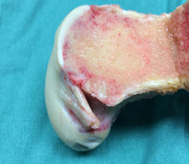
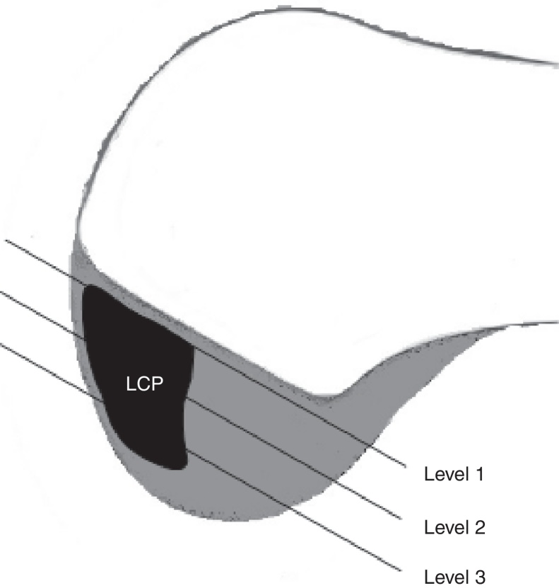
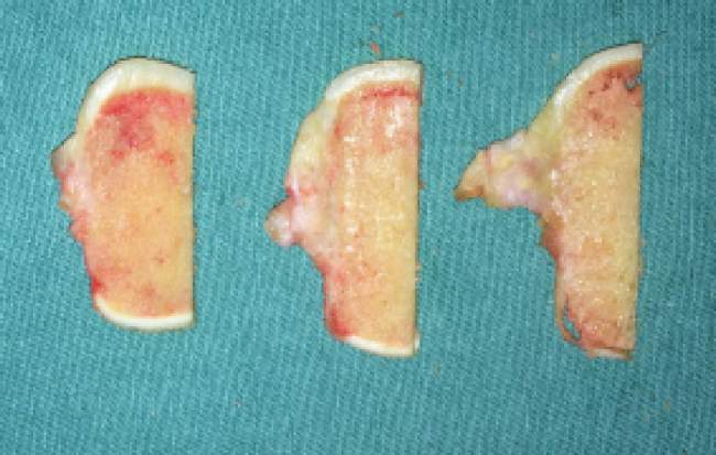
Ten cadaveric knees were used for this study. The posterior cruciate ligament femoral insertion was observed microscopically. Hematoxylin and eosin staining was performed to observe the morphology of the posterior cruciate ligament insertion. Alcian blue staining was performed to determine the location of the cartilage matrix and better assist in the observation and differentiation between direct and indirect insertions.
Results
The direct insertion was observed to be a more complex structure than the indirect insertion because it showed four different histological layers (ligament, uncalcified fibrocartilage, calcified fibrocartilage, and bone). Chondrocytes were observed in the uncalcified and calcified fibrocartilage layers. It was observed that the indirect insertion was composed of two layers in which the ligament was anchored directly to the bone by collagen fibers. Indirect insertion was located in the marginal region of the posterior cruciate ligament between the direct insertion and the anterior articular cartilage.
Conclusion
Through histological analysis, it was demonstrated that the indirect insertion was adjacent to the anterior articular cartilage and presents a histological pattern where the collagen fibers insert directly into the bone (two-layer insertion). The direct insertion is posterior to the indirect insertion and has four histologically distinct layers.

 求助内容:
求助内容: 应助结果提醒方式:


