Diego da Costa Astur, João Victor Novaretti, Andre Cicone Liggieri, César Janovsky, Alexandre Pedro Nicolini, Moises Cohen
{"title":"超声评估腘绳肌腱直径:能否预测移植物的大小?","authors":"Diego da Costa Astur, João Victor Novaretti, Andre Cicone Liggieri, César Janovsky, Alexandre Pedro Nicolini, Moises Cohen","doi":"10.1016/j.rboe.2018.05.005","DOIUrl":null,"url":null,"abstract":"<div><h3>Objective</h3><p>Perform the preoperative measurement of the hamstring tendons using ultrasound imaging, validating and correlating the measured value with that found during surgical reconstruction of the ligament.</p></div><div><h3>Methods</h3><p>A cross-sectional study was carried out with 24 patients who underwent ultrasonographic measurement of the semitendinosus and gracilis muscle tendons and were subsequently submitted to surgical reconstruction of the ACL, with ipsilateral semitendinosus and gracilis tendon grafting.</p></div><div><h3>Results</h3><p>The patients’ ages ranged from 16 to 43 years, with a mean of 24.8 years (SD<!--> <!-->=<!--> <!-->8.4 years), 79.2% were men, and the distribution by side was 41.7% right knees and 58.3% left knees. A non-significant correlation coefficient was found between the area calculated by ultrasound (2<!--> <!-->×<!--> <!-->semitendinosus area<!--> <!-->+<!--> <!-->2<!--> <!-->×<!--> <!-->gracilis area) and the intraoperative measurement (<em>r</em> <!-->=<!--> <!-->0.16; <em>p</em> <!-->=<!--> <!-->0.443). No evidence of a difference between intraoperative measurements <8<!--> <!-->mm and ≥8<!--> <!-->mm was found for the area calculated by the ultrasound (<em>p</em> <!-->=<!--> <!-->0.746). The difference observed between the groups was −0.01 (95% CI: −0.09 to 0.07).</p></div><div><h3>Conclusion</h3><p>Preoperative ultrasound imaging of the semitendinosus and gracilis tendons does not present a statistically significant correlation with the intraoperative measurement of the quadruple hamstring graft for ligament reconstruction.</p></div>","PeriodicalId":101095,"journal":{"name":"Revista Brasileira de Ortopedia (English Edition)","volume":"53 4","pages":"Pages 404-409"},"PeriodicalIF":0.0000,"publicationDate":"2018-07-01","publicationTypes":"Journal Article","fieldsOfStudy":null,"isOpenAccess":false,"openAccessPdf":"https://sci-hub-pdf.com/10.1016/j.rboe.2018.05.005","citationCount":"5","resultStr":"{\"title\":\"Ultrasonography for evaluation of hamstring tendon diameter: is it possible to predict the size of the graft?\",\"authors\":\"Diego da Costa Astur, João Victor Novaretti, Andre Cicone Liggieri, César Janovsky, Alexandre Pedro Nicolini, Moises Cohen\",\"doi\":\"10.1016/j.rboe.2018.05.005\",\"DOIUrl\":null,\"url\":null,\"abstract\":\"<div><h3>Objective</h3><p>Perform the preoperative measurement of the hamstring tendons using ultrasound imaging, validating and correlating the measured value with that found during surgical reconstruction of the ligament.</p></div><div><h3>Methods</h3><p>A cross-sectional study was carried out with 24 patients who underwent ultrasonographic measurement of the semitendinosus and gracilis muscle tendons and were subsequently submitted to surgical reconstruction of the ACL, with ipsilateral semitendinosus and gracilis tendon grafting.</p></div><div><h3>Results</h3><p>The patients’ ages ranged from 16 to 43 years, with a mean of 24.8 years (SD<!--> <!-->=<!--> <!-->8.4 years), 79.2% were men, and the distribution by side was 41.7% right knees and 58.3% left knees. A non-significant correlation coefficient was found between the area calculated by ultrasound (2<!--> <!-->×<!--> <!-->semitendinosus area<!--> <!-->+<!--> <!-->2<!--> <!-->×<!--> <!-->gracilis area) and the intraoperative measurement (<em>r</em> <!-->=<!--> <!-->0.16; <em>p</em> <!-->=<!--> <!-->0.443). No evidence of a difference between intraoperative measurements <8<!--> <!-->mm and ≥8<!--> <!-->mm was found for the area calculated by the ultrasound (<em>p</em> <!-->=<!--> <!-->0.746). The difference observed between the groups was −0.01 (95% CI: −0.09 to 0.07).</p></div><div><h3>Conclusion</h3><p>Preoperative ultrasound imaging of the semitendinosus and gracilis tendons does not present a statistically significant correlation with the intraoperative measurement of the quadruple hamstring graft for ligament reconstruction.</p></div>\",\"PeriodicalId\":101095,\"journal\":{\"name\":\"Revista Brasileira de Ortopedia (English Edition)\",\"volume\":\"53 4\",\"pages\":\"Pages 404-409\"},\"PeriodicalIF\":0.0000,\"publicationDate\":\"2018-07-01\",\"publicationTypes\":\"Journal Article\",\"fieldsOfStudy\":null,\"isOpenAccess\":false,\"openAccessPdf\":\"https://sci-hub-pdf.com/10.1016/j.rboe.2018.05.005\",\"citationCount\":\"5\",\"resultStr\":null,\"platform\":\"Semanticscholar\",\"paperid\":null,\"PeriodicalName\":\"Revista Brasileira de Ortopedia (English Edition)\",\"FirstCategoryId\":\"1085\",\"ListUrlMain\":\"https://www.sciencedirect.com/science/article/pii/S2255497118300703\",\"RegionNum\":0,\"RegionCategory\":null,\"ArticlePicture\":[],\"TitleCN\":null,\"AbstractTextCN\":null,\"PMCID\":null,\"EPubDate\":\"\",\"PubModel\":\"\",\"JCR\":\"\",\"JCRName\":\"\",\"Score\":null,\"Total\":0}","platform":"Semanticscholar","paperid":null,"PeriodicalName":"Revista Brasileira de Ortopedia (English Edition)","FirstCategoryId":"1085","ListUrlMain":"https://www.sciencedirect.com/science/article/pii/S2255497118300703","RegionNum":0,"RegionCategory":null,"ArticlePicture":[],"TitleCN":null,"AbstractTextCN":null,"PMCID":null,"EPubDate":"","PubModel":"","JCR":"","JCRName":"","Score":null,"Total":0}
Ultrasonography for evaluation of hamstring tendon diameter: is it possible to predict the size of the graft?
Objective
Perform the preoperative measurement of the hamstring tendons using ultrasound imaging, validating and correlating the measured value with that found during surgical reconstruction of the ligament.
Methods
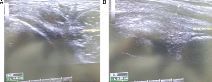
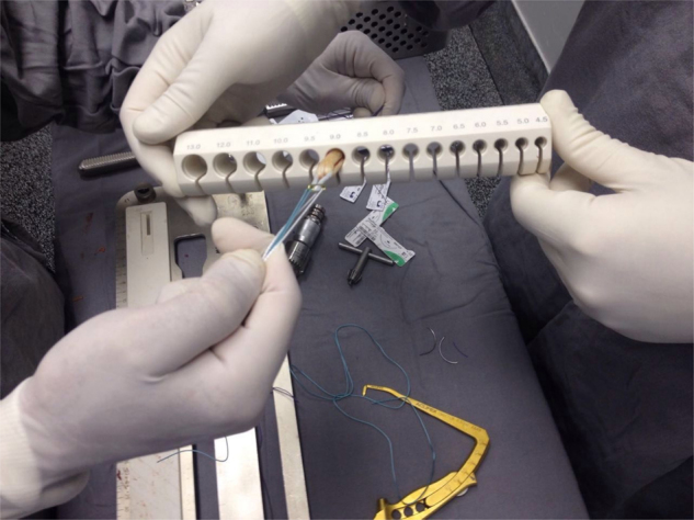
A cross-sectional study was carried out with 24 patients who underwent ultrasonographic measurement of the semitendinosus and gracilis muscle tendons and were subsequently submitted to surgical reconstruction of the ACL, with ipsilateral semitendinosus and gracilis tendon grafting.
Results
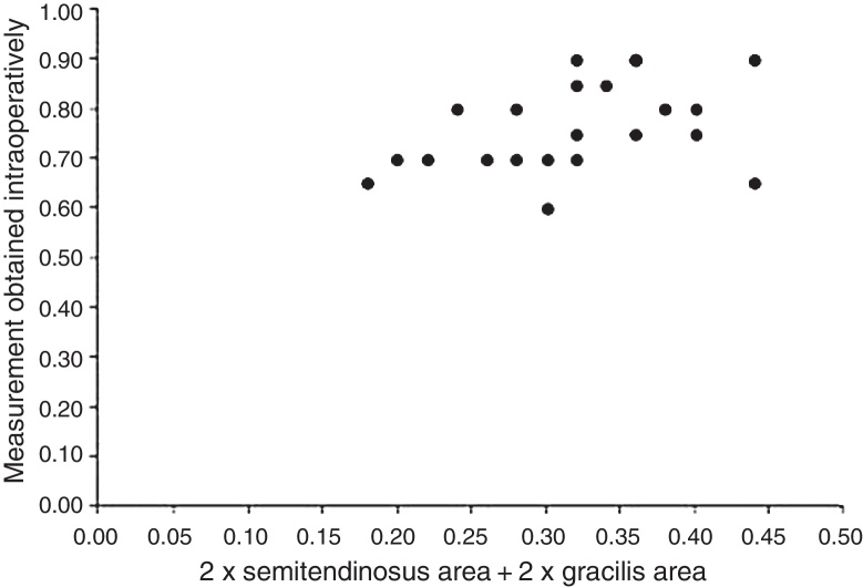
The patients’ ages ranged from 16 to 43 years, with a mean of 24.8 years (SD = 8.4 years), 79.2% were men, and the distribution by side was 41.7% right knees and 58.3% left knees. A non-significant correlation coefficient was found between the area calculated by ultrasound (2 × semitendinosus area + 2 × gracilis area) and the intraoperative measurement (r = 0.16; p = 0.443). No evidence of a difference between intraoperative measurements <8 mm and ≥8 mm was found for the area calculated by the ultrasound (p = 0.746). The difference observed between the groups was −0.01 (95% CI: −0.09 to 0.07).
Conclusion
Preoperative ultrasound imaging of the semitendinosus and gracilis tendons does not present a statistically significant correlation with the intraoperative measurement of the quadruple hamstring graft for ligament reconstruction.

 求助内容:
求助内容: 应助结果提醒方式:


