Marcello Zaia Oliveira, Mauro Batista Albano, Guilherme Augusto Stirma, Mario Massatomo Namba, Leandro Vidigal, Luiz Antonio Munhoz da Cunha
{"title":"透明质酸在实验性骨关节炎模型中的关节内粘补充","authors":"Marcello Zaia Oliveira, Mauro Batista Albano, Guilherme Augusto Stirma, Mario Massatomo Namba, Leandro Vidigal, Luiz Antonio Munhoz da Cunha","doi":"10.1016/j.rboe.2018.03.009","DOIUrl":null,"url":null,"abstract":"<div><h3>Objective</h3><p>To analyze, from the immunohistochemical perspective, the effects of hyaluronic acid of different molecular weights in an experimental model of osteoarthritis in rabbits.</p></div><div><h3>Methods</h3><p>Forty-four male California rabbits were randomly assigned to three different groups (PR, S, and P) and submitted to the resection of the anterior cruciate ligament of the right knee. Three weeks after the surgical procedure, three intra-articular weekly injections were carried out with low-molecular-weight native hyaluronic acid (Hyalgan®) to PR group, high molecular weight branched chain hyaluronic acid (Synvisc®) to group S, and saline solution 0.9% to group P. All animals were sacrificed 12 weeks after the surgical procedure, and the tibial plateaus of the infiltrated knees were then dissected. Histological sections of cartilage from the tibial plateau support areas were stained with immunohistochemical markers in order to investigate the amount of metalloproteases (MMPs 3 and 13) and their inhibitors (TIMPs 1 and 3). The staining intensity was quantified on a Zeiss Imager.Z2 Metasystems microscope and analyzed by Metafer4 Msearch software.</p></div><div><h3>Results</h3><p>The chondroprotective effect of the hyaluronic acids used in the study was demonstrated when compared to the control group. However, the comparison between them presented no significant statistical difference regarding chondroprotection.</p></div><div><h3>Conclusion</h3><p>The injection of saline solution demonstrated signs of OA development, while adding native hyaluronic acid of low molecular weight (Hyalgan®) and hyaluronic acid of high molecular weight (Synvisc®) protected the articular cartilage in this model of OA.</p></div>","PeriodicalId":101095,"journal":{"name":"Revista Brasileira de Ortopedia (English Edition)","volume":"53 3","pages":"Pages 293-299"},"PeriodicalIF":0.0000,"publicationDate":"2018-05-01","publicationTypes":"Journal Article","fieldsOfStudy":null,"isOpenAccess":false,"openAccessPdf":"https://sci-hub-pdf.com/10.1016/j.rboe.2018.03.009","citationCount":"5","resultStr":"{\"title\":\"Intra-articular viscosupplementation of hyaluronic acids in an experimental osteoarthritis model\",\"authors\":\"Marcello Zaia Oliveira, Mauro Batista Albano, Guilherme Augusto Stirma, Mario Massatomo Namba, Leandro Vidigal, Luiz Antonio Munhoz da Cunha\",\"doi\":\"10.1016/j.rboe.2018.03.009\",\"DOIUrl\":null,\"url\":null,\"abstract\":\"<div><h3>Objective</h3><p>To analyze, from the immunohistochemical perspective, the effects of hyaluronic acid of different molecular weights in an experimental model of osteoarthritis in rabbits.</p></div><div><h3>Methods</h3><p>Forty-four male California rabbits were randomly assigned to three different groups (PR, S, and P) and submitted to the resection of the anterior cruciate ligament of the right knee. Three weeks after the surgical procedure, three intra-articular weekly injections were carried out with low-molecular-weight native hyaluronic acid (Hyalgan®) to PR group, high molecular weight branched chain hyaluronic acid (Synvisc®) to group S, and saline solution 0.9% to group P. All animals were sacrificed 12 weeks after the surgical procedure, and the tibial plateaus of the infiltrated knees were then dissected. Histological sections of cartilage from the tibial plateau support areas were stained with immunohistochemical markers in order to investigate the amount of metalloproteases (MMPs 3 and 13) and their inhibitors (TIMPs 1 and 3). The staining intensity was quantified on a Zeiss Imager.Z2 Metasystems microscope and analyzed by Metafer4 Msearch software.</p></div><div><h3>Results</h3><p>The chondroprotective effect of the hyaluronic acids used in the study was demonstrated when compared to the control group. However, the comparison between them presented no significant statistical difference regarding chondroprotection.</p></div><div><h3>Conclusion</h3><p>The injection of saline solution demonstrated signs of OA development, while adding native hyaluronic acid of low molecular weight (Hyalgan®) and hyaluronic acid of high molecular weight (Synvisc®) protected the articular cartilage in this model of OA.</p></div>\",\"PeriodicalId\":101095,\"journal\":{\"name\":\"Revista Brasileira de Ortopedia (English Edition)\",\"volume\":\"53 3\",\"pages\":\"Pages 293-299\"},\"PeriodicalIF\":0.0000,\"publicationDate\":\"2018-05-01\",\"publicationTypes\":\"Journal Article\",\"fieldsOfStudy\":null,\"isOpenAccess\":false,\"openAccessPdf\":\"https://sci-hub-pdf.com/10.1016/j.rboe.2018.03.009\",\"citationCount\":\"5\",\"resultStr\":null,\"platform\":\"Semanticscholar\",\"paperid\":null,\"PeriodicalName\":\"Revista Brasileira de Ortopedia (English Edition)\",\"FirstCategoryId\":\"1085\",\"ListUrlMain\":\"https://www.sciencedirect.com/science/article/pii/S225549711830048X\",\"RegionNum\":0,\"RegionCategory\":null,\"ArticlePicture\":[],\"TitleCN\":null,\"AbstractTextCN\":null,\"PMCID\":null,\"EPubDate\":\"\",\"PubModel\":\"\",\"JCR\":\"\",\"JCRName\":\"\",\"Score\":null,\"Total\":0}","platform":"Semanticscholar","paperid":null,"PeriodicalName":"Revista Brasileira de Ortopedia (English Edition)","FirstCategoryId":"1085","ListUrlMain":"https://www.sciencedirect.com/science/article/pii/S225549711830048X","RegionNum":0,"RegionCategory":null,"ArticlePicture":[],"TitleCN":null,"AbstractTextCN":null,"PMCID":null,"EPubDate":"","PubModel":"","JCR":"","JCRName":"","Score":null,"Total":0}
Intra-articular viscosupplementation of hyaluronic acids in an experimental osteoarthritis model
Objective
To analyze, from the immunohistochemical perspective, the effects of hyaluronic acid of different molecular weights in an experimental model of osteoarthritis in rabbits.
Methods
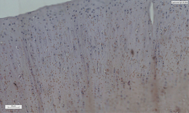
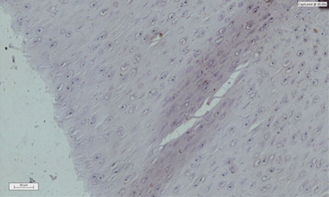
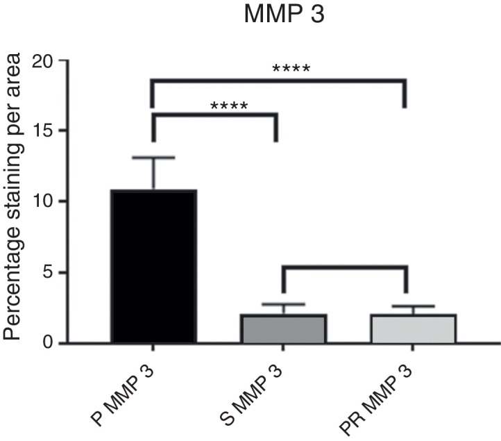
Forty-four male California rabbits were randomly assigned to three different groups (PR, S, and P) and submitted to the resection of the anterior cruciate ligament of the right knee. Three weeks after the surgical procedure, three intra-articular weekly injections were carried out with low-molecular-weight native hyaluronic acid (Hyalgan®) to PR group, high molecular weight branched chain hyaluronic acid (Synvisc®) to group S, and saline solution 0.9% to group P. All animals were sacrificed 12 weeks after the surgical procedure, and the tibial plateaus of the infiltrated knees were then dissected. Histological sections of cartilage from the tibial plateau support areas were stained with immunohistochemical markers in order to investigate the amount of metalloproteases (MMPs 3 and 13) and their inhibitors (TIMPs 1 and 3). The staining intensity was quantified on a Zeiss Imager.Z2 Metasystems microscope and analyzed by Metafer4 Msearch software.
Results
The chondroprotective effect of the hyaluronic acids used in the study was demonstrated when compared to the control group. However, the comparison between them presented no significant statistical difference regarding chondroprotection.
Conclusion
The injection of saline solution demonstrated signs of OA development, while adding native hyaluronic acid of low molecular weight (Hyalgan®) and hyaluronic acid of high molecular weight (Synvisc®) protected the articular cartilage in this model of OA.

 求助内容:
求助内容: 应助结果提醒方式:


