{"title":"正电子发射断层成像中枢神经系统脱髓鞘和再髓鞘。","authors":"Benedetta Bodini, Bruno Stankoff","doi":"10.3233/BPL-160042","DOIUrl":null,"url":null,"abstract":"<p><p>Positron Emission Tomography (PET), an imaging technique based on the injection of radiotracers directed against specific biological targets within brain tissues, within brain tissues, is a specific and sensitive technique which offers the unique opportunity to quantify myelin dynamics in the central nervous system. Several stilbene and benzothiazole derivatives have been repurposed to image myelin by PET. In demyelinating and dysmyelinating models, selected radiotracers were shown to reliably quantify demyelination and remyelination, allowing a translational approach in humans. A pilot study in subjects with active relapsing MS using PET and the most available benzothiazole derivative, [<sup>11</sup>C]PIB, supported the hypothesis that this technique is able to quantify myelin content in multiple sclerosis (MS) lesions and to capture dynamic demyelination and remyelination over time. This study highlighted for the first <i>time in vivo</i> the prognostic value of individual profiles of remyelination on the disease course. In future, the clinical application of myelin PET will be pushed forward thanks to the availability of novel fluorinated tracers for myelin, together with the setting up of non invasive quantification procedures and the use of powerful PET-MR systems. This will enable to address <i>in vivo</i> critical unanswered questions about the pathogenesis of remyelination, and to measure the efficacy of emerging promyelinating drugs in early-phase therapeutic trials.</p>","PeriodicalId":72451,"journal":{"name":"Brain plasticity (Amsterdam, Netherlands)","volume":"2 1","pages":"93-98"},"PeriodicalIF":0.0000,"publicationDate":"2016-12-21","publicationTypes":"Journal Article","fieldsOfStudy":null,"isOpenAccess":false,"openAccessPdf":"https://sci-hub-pdf.com/10.3233/BPL-160042","citationCount":"8","resultStr":"{\"title\":\"Imaging Central Nervous System Demyelination and Remyelination by Positron-Emission Tomography.\",\"authors\":\"Benedetta Bodini, Bruno Stankoff\",\"doi\":\"10.3233/BPL-160042\",\"DOIUrl\":null,\"url\":null,\"abstract\":\"<p><p>Positron Emission Tomography (PET), an imaging technique based on the injection of radiotracers directed against specific biological targets within brain tissues, within brain tissues, is a specific and sensitive technique which offers the unique opportunity to quantify myelin dynamics in the central nervous system. Several stilbene and benzothiazole derivatives have been repurposed to image myelin by PET. In demyelinating and dysmyelinating models, selected radiotracers were shown to reliably quantify demyelination and remyelination, allowing a translational approach in humans. A pilot study in subjects with active relapsing MS using PET and the most available benzothiazole derivative, [<sup>11</sup>C]PIB, supported the hypothesis that this technique is able to quantify myelin content in multiple sclerosis (MS) lesions and to capture dynamic demyelination and remyelination over time. This study highlighted for the first <i>time in vivo</i> the prognostic value of individual profiles of remyelination on the disease course. In future, the clinical application of myelin PET will be pushed forward thanks to the availability of novel fluorinated tracers for myelin, together with the setting up of non invasive quantification procedures and the use of powerful PET-MR systems. This will enable to address <i>in vivo</i> critical unanswered questions about the pathogenesis of remyelination, and to measure the efficacy of emerging promyelinating drugs in early-phase therapeutic trials.</p>\",\"PeriodicalId\":72451,\"journal\":{\"name\":\"Brain plasticity (Amsterdam, Netherlands)\",\"volume\":\"2 1\",\"pages\":\"93-98\"},\"PeriodicalIF\":0.0000,\"publicationDate\":\"2016-12-21\",\"publicationTypes\":\"Journal Article\",\"fieldsOfStudy\":null,\"isOpenAccess\":false,\"openAccessPdf\":\"https://sci-hub-pdf.com/10.3233/BPL-160042\",\"citationCount\":\"8\",\"resultStr\":null,\"platform\":\"Semanticscholar\",\"paperid\":null,\"PeriodicalName\":\"Brain plasticity (Amsterdam, Netherlands)\",\"FirstCategoryId\":\"1085\",\"ListUrlMain\":\"https://doi.org/10.3233/BPL-160042\",\"RegionNum\":0,\"RegionCategory\":null,\"ArticlePicture\":[],\"TitleCN\":null,\"AbstractTextCN\":null,\"PMCID\":null,\"EPubDate\":\"\",\"PubModel\":\"\",\"JCR\":\"\",\"JCRName\":\"\",\"Score\":null,\"Total\":0}","platform":"Semanticscholar","paperid":null,"PeriodicalName":"Brain plasticity (Amsterdam, Netherlands)","FirstCategoryId":"1085","ListUrlMain":"https://doi.org/10.3233/BPL-160042","RegionNum":0,"RegionCategory":null,"ArticlePicture":[],"TitleCN":null,"AbstractTextCN":null,"PMCID":null,"EPubDate":"","PubModel":"","JCR":"","JCRName":"","Score":null,"Total":0}
Imaging Central Nervous System Demyelination and Remyelination by Positron-Emission Tomography.
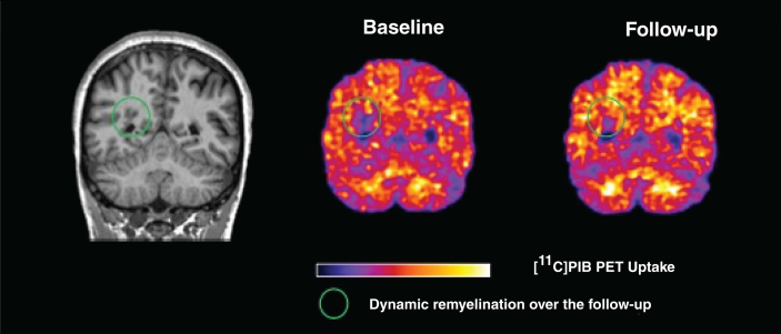
Positron Emission Tomography (PET), an imaging technique based on the injection of radiotracers directed against specific biological targets within brain tissues, within brain tissues, is a specific and sensitive technique which offers the unique opportunity to quantify myelin dynamics in the central nervous system. Several stilbene and benzothiazole derivatives have been repurposed to image myelin by PET. In demyelinating and dysmyelinating models, selected radiotracers were shown to reliably quantify demyelination and remyelination, allowing a translational approach in humans. A pilot study in subjects with active relapsing MS using PET and the most available benzothiazole derivative, [11C]PIB, supported the hypothesis that this technique is able to quantify myelin content in multiple sclerosis (MS) lesions and to capture dynamic demyelination and remyelination over time. This study highlighted for the first time in vivo the prognostic value of individual profiles of remyelination on the disease course. In future, the clinical application of myelin PET will be pushed forward thanks to the availability of novel fluorinated tracers for myelin, together with the setting up of non invasive quantification procedures and the use of powerful PET-MR systems. This will enable to address in vivo critical unanswered questions about the pathogenesis of remyelination, and to measure the efficacy of emerging promyelinating drugs in early-phase therapeutic trials.

 求助内容:
求助内容: 应助结果提醒方式:


