{"title":"脑叶下发育不良-神经影像学、脑磁图和组织病理学综合评价","authors":"Kenchaiah Raghavendra , Ganne Chaitanya , Bhargava Goutham , Anita Mahadevan , Ravindranadh Chowdary Mundlamuri , Rose Dawn Bharath , Mariyappa Narayannan , Malla Bhaskar Rao , Arimappamagan Arivazhagan , Parthasarthy Satishchandra , Sanjib Sinha","doi":"10.1016/j.ebcr.2017.11.002","DOIUrl":null,"url":null,"abstract":"<div><p>Sublobar dysplasia, a rare cortical malformation has been defined in only 8 patients to date. It was identified on the basis of histopathological features and MRI findings. We report a right temporal sublobar dysplasia, with detailed evaluation including neuroimaging, magnetoencephalography and histopathology to further characterize the pathology. Additional pathological features included a deep collateral sulcus in the basal right temporal lobe, thinned out right corticospinal tract, and bilateral asymmetric basal ganglia changes. Magnetoencephalograpy localized the seizure focus to the posterior margin of the dysplasia. Histopathological evaluation helped exclude other types of dysplasia. Similar to a previous study, the child had Engel 1a outcome.</p></div>","PeriodicalId":56365,"journal":{"name":"Epilepsy and Behavior Case Reports","volume":"9 ","pages":"Pages 22-25"},"PeriodicalIF":0.0000,"publicationDate":"2018-01-01","publicationTypes":"Journal Article","fieldsOfStudy":null,"isOpenAccess":false,"openAccessPdf":"https://sci-hub-pdf.com/10.1016/j.ebcr.2017.11.002","citationCount":"2","resultStr":"{\"title\":\"Sub-lobar dysplasia — A comprehensive evaluation with neuroimaging, magnetoencephalography and histopathology\",\"authors\":\"Kenchaiah Raghavendra , Ganne Chaitanya , Bhargava Goutham , Anita Mahadevan , Ravindranadh Chowdary Mundlamuri , Rose Dawn Bharath , Mariyappa Narayannan , Malla Bhaskar Rao , Arimappamagan Arivazhagan , Parthasarthy Satishchandra , Sanjib Sinha\",\"doi\":\"10.1016/j.ebcr.2017.11.002\",\"DOIUrl\":null,\"url\":null,\"abstract\":\"<div><p>Sublobar dysplasia, a rare cortical malformation has been defined in only 8 patients to date. It was identified on the basis of histopathological features and MRI findings. We report a right temporal sublobar dysplasia, with detailed evaluation including neuroimaging, magnetoencephalography and histopathology to further characterize the pathology. Additional pathological features included a deep collateral sulcus in the basal right temporal lobe, thinned out right corticospinal tract, and bilateral asymmetric basal ganglia changes. Magnetoencephalograpy localized the seizure focus to the posterior margin of the dysplasia. Histopathological evaluation helped exclude other types of dysplasia. Similar to a previous study, the child had Engel 1a outcome.</p></div>\",\"PeriodicalId\":56365,\"journal\":{\"name\":\"Epilepsy and Behavior Case Reports\",\"volume\":\"9 \",\"pages\":\"Pages 22-25\"},\"PeriodicalIF\":0.0000,\"publicationDate\":\"2018-01-01\",\"publicationTypes\":\"Journal Article\",\"fieldsOfStudy\":null,\"isOpenAccess\":false,\"openAccessPdf\":\"https://sci-hub-pdf.com/10.1016/j.ebcr.2017.11.002\",\"citationCount\":\"2\",\"resultStr\":null,\"platform\":\"Semanticscholar\",\"paperid\":null,\"PeriodicalName\":\"Epilepsy and Behavior Case Reports\",\"FirstCategoryId\":\"1085\",\"ListUrlMain\":\"https://www.sciencedirect.com/science/article/pii/S2213323217301196\",\"RegionNum\":0,\"RegionCategory\":null,\"ArticlePicture\":[],\"TitleCN\":null,\"AbstractTextCN\":null,\"PMCID\":null,\"EPubDate\":\"\",\"PubModel\":\"\",\"JCR\":\"\",\"JCRName\":\"\",\"Score\":null,\"Total\":0}","platform":"Semanticscholar","paperid":null,"PeriodicalName":"Epilepsy and Behavior Case Reports","FirstCategoryId":"1085","ListUrlMain":"https://www.sciencedirect.com/science/article/pii/S2213323217301196","RegionNum":0,"RegionCategory":null,"ArticlePicture":[],"TitleCN":null,"AbstractTextCN":null,"PMCID":null,"EPubDate":"","PubModel":"","JCR":"","JCRName":"","Score":null,"Total":0}
Sub-lobar dysplasia — A comprehensive evaluation with neuroimaging, magnetoencephalography and histopathology
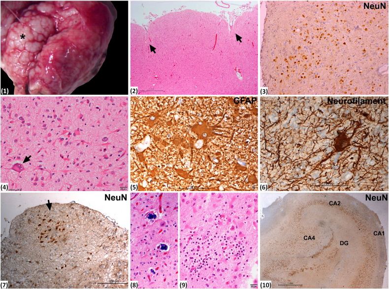
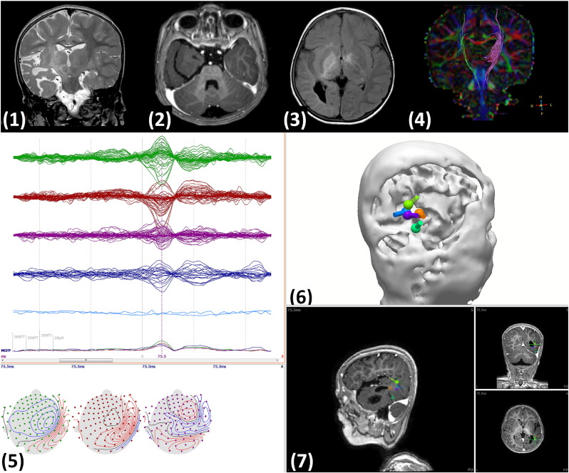
Sublobar dysplasia, a rare cortical malformation has been defined in only 8 patients to date. It was identified on the basis of histopathological features and MRI findings. We report a right temporal sublobar dysplasia, with detailed evaluation including neuroimaging, magnetoencephalography and histopathology to further characterize the pathology. Additional pathological features included a deep collateral sulcus in the basal right temporal lobe, thinned out right corticospinal tract, and bilateral asymmetric basal ganglia changes. Magnetoencephalograpy localized the seizure focus to the posterior margin of the dysplasia. Histopathological evaluation helped exclude other types of dysplasia. Similar to a previous study, the child had Engel 1a outcome.

 求助内容:
求助内容: 应助结果提醒方式:


