Dong Hwan Kim, Dong Ha Kim, Kyoung Hyup Nam, Byung Kwan Choi, In Ho Han
{"title":"颈椎自发性硬膜外血肿伴静脉静脉结石及其可能的发病机制。","authors":"Dong Hwan Kim, Dong Ha Kim, Kyoung Hyup Nam, Byung Kwan Choi, In Ho Han","doi":"10.14245/kjs.2017.14.3.96","DOIUrl":null,"url":null,"abstract":"<p><p>Although the etiology of spontaneous spinal epidural hematoma (SSEH) is unclear, SSEH is known to be associated with anticoagulants, coagulopathy, vascular malformation, hypertension, and pregnancy. However, no report has been issued on the relation between SSEH and venous phlebolith. Here, the authors present an extremely rare case of SSEH associated with phlebolith in the cervical spine and suggest a possible pathogenesis. A 36-year-old man without any relevant medical history presented with neck pain and numbness and severe radiating pain on the left arm. Magnetic resonance imaging showed epidural hematoma at the C5-7 level, and computed tomography revealed a calcified nodule in the left epidural hemorrhage at C6 level. During left partial laminectomy, epidural venous plexus, and thick epidural hematoma were found, and hematoma removal revealed a white, ovoid, smooth, hard mass of diameter 3 mm. Histopathologic examination confirmed the mass as a venous phlebolith. The presence of a calcified solitary nodule in dorsal epidural space indicates the presence of phlebolith and the risk of SSEH. In such cases, the authors recommend spine surgeons should take into consideration the possibility of epidural hemorrhage.</p>","PeriodicalId":17867,"journal":{"name":"Korean Journal of Spine","volume":"14 3","pages":"96-98"},"PeriodicalIF":0.0000,"publicationDate":"2017-09-01","publicationTypes":"Journal Article","fieldsOfStudy":null,"isOpenAccess":false,"openAccessPdf":"https://ftp.ncbi.nlm.nih.gov/pub/pmc/oa_pdf/5e/35/kjs-14-3-96.PMC5642097.pdf","citationCount":"0","resultStr":"{\"title\":\"Spontaneous Epidural Hematoma Associated with Venous Phlebolith in Cervical Spine and Possible Pathogenesis.\",\"authors\":\"Dong Hwan Kim, Dong Ha Kim, Kyoung Hyup Nam, Byung Kwan Choi, In Ho Han\",\"doi\":\"10.14245/kjs.2017.14.3.96\",\"DOIUrl\":null,\"url\":null,\"abstract\":\"<p><p>Although the etiology of spontaneous spinal epidural hematoma (SSEH) is unclear, SSEH is known to be associated with anticoagulants, coagulopathy, vascular malformation, hypertension, and pregnancy. However, no report has been issued on the relation between SSEH and venous phlebolith. Here, the authors present an extremely rare case of SSEH associated with phlebolith in the cervical spine and suggest a possible pathogenesis. A 36-year-old man without any relevant medical history presented with neck pain and numbness and severe radiating pain on the left arm. Magnetic resonance imaging showed epidural hematoma at the C5-7 level, and computed tomography revealed a calcified nodule in the left epidural hemorrhage at C6 level. During left partial laminectomy, epidural venous plexus, and thick epidural hematoma were found, and hematoma removal revealed a white, ovoid, smooth, hard mass of diameter 3 mm. Histopathologic examination confirmed the mass as a venous phlebolith. The presence of a calcified solitary nodule in dorsal epidural space indicates the presence of phlebolith and the risk of SSEH. In such cases, the authors recommend spine surgeons should take into consideration the possibility of epidural hemorrhage.</p>\",\"PeriodicalId\":17867,\"journal\":{\"name\":\"Korean Journal of Spine\",\"volume\":\"14 3\",\"pages\":\"96-98\"},\"PeriodicalIF\":0.0000,\"publicationDate\":\"2017-09-01\",\"publicationTypes\":\"Journal Article\",\"fieldsOfStudy\":null,\"isOpenAccess\":false,\"openAccessPdf\":\"https://ftp.ncbi.nlm.nih.gov/pub/pmc/oa_pdf/5e/35/kjs-14-3-96.PMC5642097.pdf\",\"citationCount\":\"0\",\"resultStr\":null,\"platform\":\"Semanticscholar\",\"paperid\":null,\"PeriodicalName\":\"Korean Journal of Spine\",\"FirstCategoryId\":\"1085\",\"ListUrlMain\":\"https://doi.org/10.14245/kjs.2017.14.3.96\",\"RegionNum\":0,\"RegionCategory\":null,\"ArticlePicture\":[],\"TitleCN\":null,\"AbstractTextCN\":null,\"PMCID\":null,\"EPubDate\":\"2017/9/30 0:00:00\",\"PubModel\":\"Epub\",\"JCR\":\"\",\"JCRName\":\"\",\"Score\":null,\"Total\":0}","platform":"Semanticscholar","paperid":null,"PeriodicalName":"Korean Journal of Spine","FirstCategoryId":"1085","ListUrlMain":"https://doi.org/10.14245/kjs.2017.14.3.96","RegionNum":0,"RegionCategory":null,"ArticlePicture":[],"TitleCN":null,"AbstractTextCN":null,"PMCID":null,"EPubDate":"2017/9/30 0:00:00","PubModel":"Epub","JCR":"","JCRName":"","Score":null,"Total":0}
Spontaneous Epidural Hematoma Associated with Venous Phlebolith in Cervical Spine and Possible Pathogenesis.
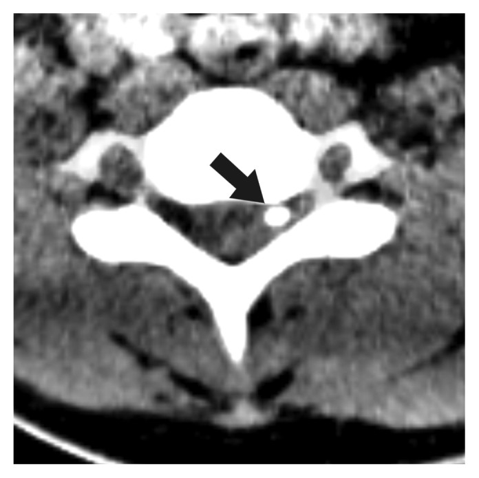
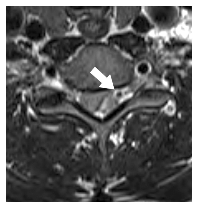
Although the etiology of spontaneous spinal epidural hematoma (SSEH) is unclear, SSEH is known to be associated with anticoagulants, coagulopathy, vascular malformation, hypertension, and pregnancy. However, no report has been issued on the relation between SSEH and venous phlebolith. Here, the authors present an extremely rare case of SSEH associated with phlebolith in the cervical spine and suggest a possible pathogenesis. A 36-year-old man without any relevant medical history presented with neck pain and numbness and severe radiating pain on the left arm. Magnetic resonance imaging showed epidural hematoma at the C5-7 level, and computed tomography revealed a calcified nodule in the left epidural hemorrhage at C6 level. During left partial laminectomy, epidural venous plexus, and thick epidural hematoma were found, and hematoma removal revealed a white, ovoid, smooth, hard mass of diameter 3 mm. Histopathologic examination confirmed the mass as a venous phlebolith. The presence of a calcified solitary nodule in dorsal epidural space indicates the presence of phlebolith and the risk of SSEH. In such cases, the authors recommend spine surgeons should take into consideration the possibility of epidural hemorrhage.

 求助内容:
求助内容: 应助结果提醒方式:


