{"title":"锥束计算机断层扫描检测上颌四根第二磨牙的治疗。","authors":"Nahid Mohammadzade Akhlaghi, Mahta Fazlyab","doi":"","DOIUrl":null,"url":null,"abstract":"<p><p>The significance of clinician's knowledge about root canal anatomy and its possible variations cannot be overlooked. In some cases, taking advantage of complementary imaging techniques can help achieve a perfect flawless endodontic treatment. This article reports endodontic management of a second maxillary molar that had an uncommon anatomy of the chamber floor. After obtaining a cone-beam computed tomography (CBCT) image, the presence of a second palatal root was confirmed. All four roots were treated and patient's symptoms were resolved.</p>","PeriodicalId":30286,"journal":{"name":"Journal of Dentistry of Tehran University of Medical Sciences","volume":"14 2","pages":"100-104"},"PeriodicalIF":0.0000,"publicationDate":"2017-03-01","publicationTypes":"Journal Article","fieldsOfStudy":null,"isOpenAccess":false,"openAccessPdf":"https://www.ncbi.nlm.nih.gov/pmc/articles/PMC5662507/pdf/","citationCount":"0","resultStr":"{\"title\":\"Treatment of a Four-Rooted Maxillary Second Molar Detected with Cone-Beam Computed Tomography.\",\"authors\":\"Nahid Mohammadzade Akhlaghi, Mahta Fazlyab\",\"doi\":\"\",\"DOIUrl\":null,\"url\":null,\"abstract\":\"<p><p>The significance of clinician's knowledge about root canal anatomy and its possible variations cannot be overlooked. In some cases, taking advantage of complementary imaging techniques can help achieve a perfect flawless endodontic treatment. This article reports endodontic management of a second maxillary molar that had an uncommon anatomy of the chamber floor. After obtaining a cone-beam computed tomography (CBCT) image, the presence of a second palatal root was confirmed. All four roots were treated and patient's symptoms were resolved.</p>\",\"PeriodicalId\":30286,\"journal\":{\"name\":\"Journal of Dentistry of Tehran University of Medical Sciences\",\"volume\":\"14 2\",\"pages\":\"100-104\"},\"PeriodicalIF\":0.0000,\"publicationDate\":\"2017-03-01\",\"publicationTypes\":\"Journal Article\",\"fieldsOfStudy\":null,\"isOpenAccess\":false,\"openAccessPdf\":\"https://www.ncbi.nlm.nih.gov/pmc/articles/PMC5662507/pdf/\",\"citationCount\":\"0\",\"resultStr\":null,\"platform\":\"Semanticscholar\",\"paperid\":null,\"PeriodicalName\":\"Journal of Dentistry of Tehran University of Medical Sciences\",\"FirstCategoryId\":\"1085\",\"ListUrlMain\":\"\",\"RegionNum\":0,\"RegionCategory\":null,\"ArticlePicture\":[],\"TitleCN\":null,\"AbstractTextCN\":null,\"PMCID\":null,\"EPubDate\":\"\",\"PubModel\":\"\",\"JCR\":\"\",\"JCRName\":\"\",\"Score\":null,\"Total\":0}","platform":"Semanticscholar","paperid":null,"PeriodicalName":"Journal of Dentistry of Tehran University of Medical Sciences","FirstCategoryId":"1085","ListUrlMain":"","RegionNum":0,"RegionCategory":null,"ArticlePicture":[],"TitleCN":null,"AbstractTextCN":null,"PMCID":null,"EPubDate":"","PubModel":"","JCR":"","JCRName":"","Score":null,"Total":0}
Treatment of a Four-Rooted Maxillary Second Molar Detected with Cone-Beam Computed Tomography.
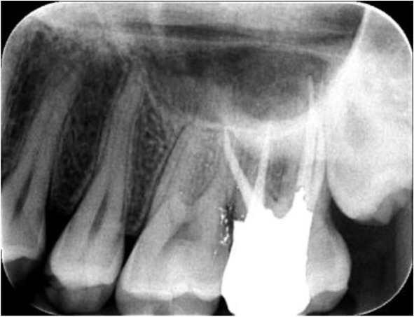
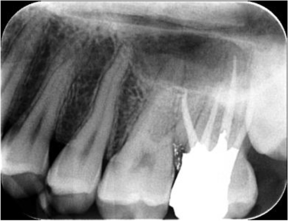
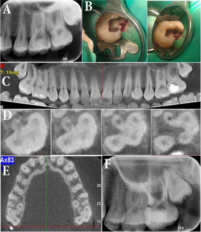
The significance of clinician's knowledge about root canal anatomy and its possible variations cannot be overlooked. In some cases, taking advantage of complementary imaging techniques can help achieve a perfect flawless endodontic treatment. This article reports endodontic management of a second maxillary molar that had an uncommon anatomy of the chamber floor. After obtaining a cone-beam computed tomography (CBCT) image, the presence of a second palatal root was confirmed. All four roots were treated and patient's symptoms were resolved.

 求助内容:
求助内容: 应助结果提醒方式:


