{"title":"基于多尺度局部基本图像特征直方图的相衬显微镜图像分割。","authors":"N Jaccard, N Szita, L D Griffin","doi":"10.1080/21681163.2015.1016243","DOIUrl":null,"url":null,"abstract":"<p><p>Phase contrast microscopy (PCM) is routinely used for the inspection of adherent cell cultures in all fields of biology and biomedicine. Key decisions for experimental protocols are often taken by an operator based on typically qualitative observations. However, automated processing and analysis of PCM images remain challenging due to the low contrast between foreground objects (cells) and background as well as various imaging artefacts. We propose a trainable pixel-wise segmentation approach whereby image structures and symmetries are encoded in the form of multi-scale Basic Image Features local histograms, and classification of them is learned by random decision trees. This approach was validated for segmentation of cell versus background, and discrimination between two different cell types. Performance close to that of state-of-the-art specialised algorithms was achieved despite the general nature of the method. The low processing time ( < 4 s per 1280 × 960 pixel images) is suitable for batch processing of experimental data as well as for interactive segmentation applications.</p>","PeriodicalId":51800,"journal":{"name":"Computer Methods in Biomechanics and Biomedical Engineering-Imaging and Visualization","volume":"5 5","pages":"359-367"},"PeriodicalIF":1.3000,"publicationDate":"2017-09-03","publicationTypes":"Journal Article","fieldsOfStudy":null,"isOpenAccess":false,"openAccessPdf":"https://www.ncbi.nlm.nih.gov/pmc/articles/PMC5526147/pdf/","citationCount":"0","resultStr":"{\"title\":\"Segmentation of phase contrast microscopy images based on multi-scale local Basic Image Features histograms.\",\"authors\":\"N Jaccard, N Szita, L D Griffin\",\"doi\":\"10.1080/21681163.2015.1016243\",\"DOIUrl\":null,\"url\":null,\"abstract\":\"<p><p>Phase contrast microscopy (PCM) is routinely used for the inspection of adherent cell cultures in all fields of biology and biomedicine. Key decisions for experimental protocols are often taken by an operator based on typically qualitative observations. However, automated processing and analysis of PCM images remain challenging due to the low contrast between foreground objects (cells) and background as well as various imaging artefacts. We propose a trainable pixel-wise segmentation approach whereby image structures and symmetries are encoded in the form of multi-scale Basic Image Features local histograms, and classification of them is learned by random decision trees. This approach was validated for segmentation of cell versus background, and discrimination between two different cell types. Performance close to that of state-of-the-art specialised algorithms was achieved despite the general nature of the method. The low processing time ( < 4 s per 1280 × 960 pixel images) is suitable for batch processing of experimental data as well as for interactive segmentation applications.</p>\",\"PeriodicalId\":51800,\"journal\":{\"name\":\"Computer Methods in Biomechanics and Biomedical Engineering-Imaging and Visualization\",\"volume\":\"5 5\",\"pages\":\"359-367\"},\"PeriodicalIF\":1.3000,\"publicationDate\":\"2017-09-03\",\"publicationTypes\":\"Journal Article\",\"fieldsOfStudy\":null,\"isOpenAccess\":false,\"openAccessPdf\":\"https://www.ncbi.nlm.nih.gov/pmc/articles/PMC5526147/pdf/\",\"citationCount\":\"0\",\"resultStr\":null,\"platform\":\"Semanticscholar\",\"paperid\":null,\"PeriodicalName\":\"Computer Methods in Biomechanics and Biomedical Engineering-Imaging and Visualization\",\"FirstCategoryId\":\"1085\",\"ListUrlMain\":\"https://doi.org/10.1080/21681163.2015.1016243\",\"RegionNum\":0,\"RegionCategory\":null,\"ArticlePicture\":[],\"TitleCN\":null,\"AbstractTextCN\":null,\"PMCID\":null,\"EPubDate\":\"2017/4/7 0:00:00\",\"PubModel\":\"Epub\",\"JCR\":\"Q4\",\"JCRName\":\"ENGINEERING, BIOMEDICAL\",\"Score\":null,\"Total\":0}","platform":"Semanticscholar","paperid":null,"PeriodicalName":"Computer Methods in Biomechanics and Biomedical Engineering-Imaging and Visualization","FirstCategoryId":"1085","ListUrlMain":"https://doi.org/10.1080/21681163.2015.1016243","RegionNum":0,"RegionCategory":null,"ArticlePicture":[],"TitleCN":null,"AbstractTextCN":null,"PMCID":null,"EPubDate":"2017/4/7 0:00:00","PubModel":"Epub","JCR":"Q4","JCRName":"ENGINEERING, BIOMEDICAL","Score":null,"Total":0}
Segmentation of phase contrast microscopy images based on multi-scale local Basic Image Features histograms.
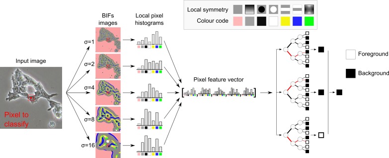
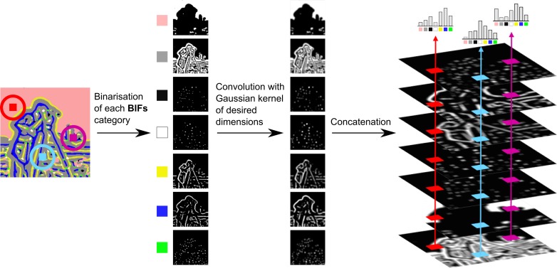
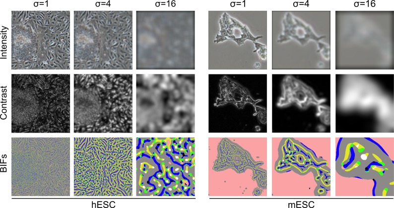
Phase contrast microscopy (PCM) is routinely used for the inspection of adherent cell cultures in all fields of biology and biomedicine. Key decisions for experimental protocols are often taken by an operator based on typically qualitative observations. However, automated processing and analysis of PCM images remain challenging due to the low contrast between foreground objects (cells) and background as well as various imaging artefacts. We propose a trainable pixel-wise segmentation approach whereby image structures and symmetries are encoded in the form of multi-scale Basic Image Features local histograms, and classification of them is learned by random decision trees. This approach was validated for segmentation of cell versus background, and discrimination between two different cell types. Performance close to that of state-of-the-art specialised algorithms was achieved despite the general nature of the method. The low processing time ( < 4 s per 1280 × 960 pixel images) is suitable for batch processing of experimental data as well as for interactive segmentation applications.
期刊介绍:
Computer Methods in Biomechanics and Biomedical Engineering: Imaging & Visualization is an international journal whose main goals are to promote solutions of excellence for both imaging and visualization of biomedical data, and establish links among researchers, clinicians, the medical technology sector and end-users. The journal provides a comprehensive forum for discussion of the current state-of-the-art in the scientific fields related to imaging and visualization, including, but not limited to: Applications of Imaging and Visualization Computational Bio- imaging and Visualization Computer Aided Diagnosis, Surgery, Therapy and Treatment Data Processing and Analysis Devices for Imaging and Visualization Grid and High Performance Computing for Imaging and Visualization Human Perception in Imaging and Visualization Image Processing and Analysis Image-based Geometric Modelling Imaging and Visualization in Biomechanics Imaging and Visualization in Biomedical Engineering Medical Clinics Medical Imaging and Visualization Multi-modal Imaging and Visualization Multiscale Imaging and Visualization Scientific Visualization Software Development for Imaging and Visualization Telemedicine Systems and Applications Virtual Reality Visual Data Mining and Knowledge Discovery.

 求助内容:
求助内容: 应助结果提醒方式:


