John Mokua Mose, David Muchina Kamau, John Maina Kagira, Naomi Maina, Maina Ngotho, Adele Njuguna, Simon Muturi Karanja
{"title":"利用从肯尼亚鸡体内分离的弓形虫开发弓形虫病神经小鼠模型。","authors":"John Mokua Mose, David Muchina Kamau, John Maina Kagira, Naomi Maina, Maina Ngotho, Adele Njuguna, Simon Muturi Karanja","doi":"10.1155/2017/4302459","DOIUrl":null,"url":null,"abstract":"<p><p>Animal models for the toxoplasmosis are scarce and have limitations. In this study, a neurological mouse model was developed in BALB/c mice infected intraperitoneally with 15 cysts of a <i>Toxoplasma gondii</i> isolate. The mice were monitored for 42 days and euthanized at different time points. Another group of mice were orally treated with dexamethasone (DXM: 2.66 mg/kg daily, 5.32 mg/kg daily) at 42 days after infection and monitored for a further 42 days. A mortality rate of 15% and 28.6% was observed in mice given 2.66 mg/kg/day and 5.32 mg/kg/day of DXM, respectively. The mean cyst numbers in the brain of DXM treated mice increased up to twofold compared with chronically infected untreated mice. Infections up to 42 days were associated with an increase in both IgM and IgG levels but following dexamethasone treatment, IgM levels declined but IgG levels continued on rising. The brain of toxoplasmosis infected mice showed mononuclear cellular infiltrations, neuronal necrosis, and cuffing. The severity of pathology was higher in mice treated with dexamethasone compared to the positive control groups. The findings of this study demonstrate that DXM-induced reactivation of chronic toxoplasmosis may be a useful development of laboratory animal model in outbred mice used for <i>in vivo</i> studies.</p>","PeriodicalId":89212,"journal":{"name":"Pathology research international","volume":"2017 ","pages":"4302459"},"PeriodicalIF":0.0000,"publicationDate":"2017-01-01","publicationTypes":"Journal Article","fieldsOfStudy":null,"isOpenAccess":false,"openAccessPdf":"https://www.ncbi.nlm.nih.gov/pmc/articles/PMC5463144/pdf/","citationCount":"0","resultStr":"{\"title\":\"Development of Neurological Mouse Model for Toxoplasmosis Using <i>Toxoplasma gondii</i> Isolated from Chicken in Kenya.\",\"authors\":\"John Mokua Mose, David Muchina Kamau, John Maina Kagira, Naomi Maina, Maina Ngotho, Adele Njuguna, Simon Muturi Karanja\",\"doi\":\"10.1155/2017/4302459\",\"DOIUrl\":null,\"url\":null,\"abstract\":\"<p><p>Animal models for the toxoplasmosis are scarce and have limitations. In this study, a neurological mouse model was developed in BALB/c mice infected intraperitoneally with 15 cysts of a <i>Toxoplasma gondii</i> isolate. The mice were monitored for 42 days and euthanized at different time points. Another group of mice were orally treated with dexamethasone (DXM: 2.66 mg/kg daily, 5.32 mg/kg daily) at 42 days after infection and monitored for a further 42 days. A mortality rate of 15% and 28.6% was observed in mice given 2.66 mg/kg/day and 5.32 mg/kg/day of DXM, respectively. The mean cyst numbers in the brain of DXM treated mice increased up to twofold compared with chronically infected untreated mice. Infections up to 42 days were associated with an increase in both IgM and IgG levels but following dexamethasone treatment, IgM levels declined but IgG levels continued on rising. The brain of toxoplasmosis infected mice showed mononuclear cellular infiltrations, neuronal necrosis, and cuffing. The severity of pathology was higher in mice treated with dexamethasone compared to the positive control groups. The findings of this study demonstrate that DXM-induced reactivation of chronic toxoplasmosis may be a useful development of laboratory animal model in outbred mice used for <i>in vivo</i> studies.</p>\",\"PeriodicalId\":89212,\"journal\":{\"name\":\"Pathology research international\",\"volume\":\"2017 \",\"pages\":\"4302459\"},\"PeriodicalIF\":0.0000,\"publicationDate\":\"2017-01-01\",\"publicationTypes\":\"Journal Article\",\"fieldsOfStudy\":null,\"isOpenAccess\":false,\"openAccessPdf\":\"https://www.ncbi.nlm.nih.gov/pmc/articles/PMC5463144/pdf/\",\"citationCount\":\"0\",\"resultStr\":null,\"platform\":\"Semanticscholar\",\"paperid\":null,\"PeriodicalName\":\"Pathology research international\",\"FirstCategoryId\":\"1085\",\"ListUrlMain\":\"https://doi.org/10.1155/2017/4302459\",\"RegionNum\":0,\"RegionCategory\":null,\"ArticlePicture\":[],\"TitleCN\":null,\"AbstractTextCN\":null,\"PMCID\":null,\"EPubDate\":\"2017/5/24 0:00:00\",\"PubModel\":\"Epub\",\"JCR\":\"\",\"JCRName\":\"\",\"Score\":null,\"Total\":0}","platform":"Semanticscholar","paperid":null,"PeriodicalName":"Pathology research international","FirstCategoryId":"1085","ListUrlMain":"https://doi.org/10.1155/2017/4302459","RegionNum":0,"RegionCategory":null,"ArticlePicture":[],"TitleCN":null,"AbstractTextCN":null,"PMCID":null,"EPubDate":"2017/5/24 0:00:00","PubModel":"Epub","JCR":"","JCRName":"","Score":null,"Total":0}
Development of Neurological Mouse Model for Toxoplasmosis Using Toxoplasma gondii Isolated from Chicken in Kenya.
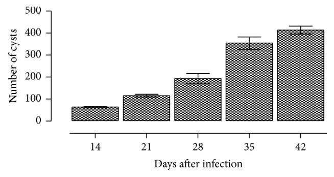
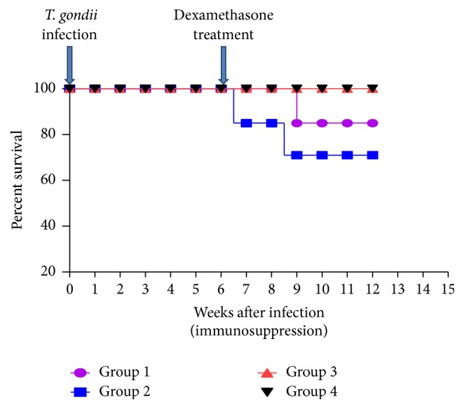
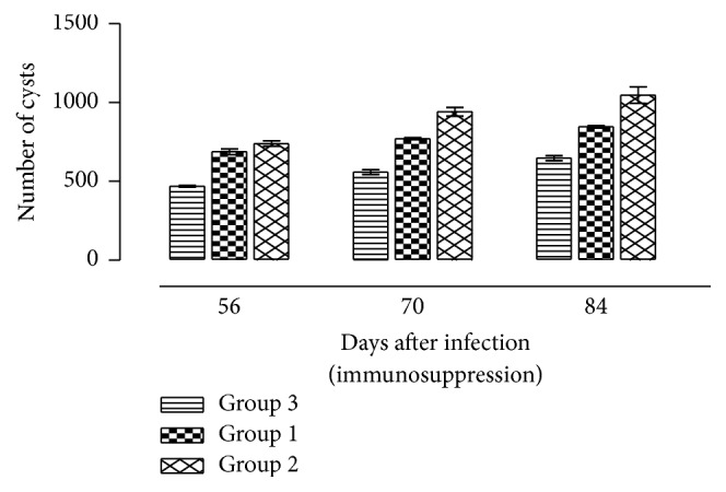
Animal models for the toxoplasmosis are scarce and have limitations. In this study, a neurological mouse model was developed in BALB/c mice infected intraperitoneally with 15 cysts of a Toxoplasma gondii isolate. The mice were monitored for 42 days and euthanized at different time points. Another group of mice were orally treated with dexamethasone (DXM: 2.66 mg/kg daily, 5.32 mg/kg daily) at 42 days after infection and monitored for a further 42 days. A mortality rate of 15% and 28.6% was observed in mice given 2.66 mg/kg/day and 5.32 mg/kg/day of DXM, respectively. The mean cyst numbers in the brain of DXM treated mice increased up to twofold compared with chronically infected untreated mice. Infections up to 42 days were associated with an increase in both IgM and IgG levels but following dexamethasone treatment, IgM levels declined but IgG levels continued on rising. The brain of toxoplasmosis infected mice showed mononuclear cellular infiltrations, neuronal necrosis, and cuffing. The severity of pathology was higher in mice treated with dexamethasone compared to the positive control groups. The findings of this study demonstrate that DXM-induced reactivation of chronic toxoplasmosis may be a useful development of laboratory animal model in outbred mice used for in vivo studies.

 求助内容:
求助内容: 应助结果提醒方式:


