{"title":"正畸牙齿移动过程中牙周变化的超声评价研究进展中。","authors":"Adela Zimbran, Diana Dudea, Cristina Gasparik, Sorin Dudea","doi":"10.15386/cjmed-663","DOIUrl":null,"url":null,"abstract":"<p><strong>Background and aim: </strong>Orthodontic tooth movement (OTM) is a process whereby the application of a force induces bone resorption on the pressure side and bone apposition on the tension side of the lamina dura. However, only limited data are available on the in vivo behavior of the periodontal tissues. The aim of this study was to assess the changes of periodontal tissues, induced by the orthodontic canine retraction, using 40 MHz ultrasonography.</p><p><strong>Methods: </strong>Ultrasonographic evaluation of periodontal tissues was conducted in 5 patients with indication for orthodontic treatment. The upper first premolars were extracted bilaterally due to severe crowding, and the canines were distalized using elastomeric chain with a net force of 100 cN. Ultrasonographic scans (US scans) were performed before, during and after retraction, in three distinct areas of the canines buccal surface: mesial, middle and distal. The reference point was the bracket, which appeared hyperechoic on the US scan. Four different dimensions were obtained: D1 (depth of the sulcus), D2 (thickness of the gingiva), D3 (length of the supracrestal fibers), D4 (width of periodontal space).</p><p><strong>Results: </strong>An increase of D1 was observed in all three areas of the periodontium, during orthodontic treatment. D3 was strongly correlated before and immediately after force delivery only for the mesial area (r=0.828, p<0.05). In total, 228 variables were statistically analyzed using Pearson's correlation coefficients, in order to demonstrate the relationship between periodontal findings during orthodontic tooth movement.</p><p><strong>Conclusion: </strong>High-resolution ultrasonography has the capability to obviate changes in periodontal ligament space and free gingiva during orthodontic tooth movement.</p>","PeriodicalId":91233,"journal":{"name":"Clujul medical (1957)","volume":"90 1","pages":"93-98"},"PeriodicalIF":0.0000,"publicationDate":"2017-01-01","publicationTypes":"Journal Article","fieldsOfStudy":null,"isOpenAccess":false,"openAccessPdf":"https://ftp.ncbi.nlm.nih.gov/pub/pmc/oa_pdf/d7/25/cm-90-93.PMC5305094.pdf","citationCount":"5","resultStr":"{\"title\":\"Ultrasonographic evaluation of periodontal changes during orthodontic tooth movement - work in progress.\",\"authors\":\"Adela Zimbran, Diana Dudea, Cristina Gasparik, Sorin Dudea\",\"doi\":\"10.15386/cjmed-663\",\"DOIUrl\":null,\"url\":null,\"abstract\":\"<p><strong>Background and aim: </strong>Orthodontic tooth movement (OTM) is a process whereby the application of a force induces bone resorption on the pressure side and bone apposition on the tension side of the lamina dura. However, only limited data are available on the in vivo behavior of the periodontal tissues. The aim of this study was to assess the changes of periodontal tissues, induced by the orthodontic canine retraction, using 40 MHz ultrasonography.</p><p><strong>Methods: </strong>Ultrasonographic evaluation of periodontal tissues was conducted in 5 patients with indication for orthodontic treatment. The upper first premolars were extracted bilaterally due to severe crowding, and the canines were distalized using elastomeric chain with a net force of 100 cN. Ultrasonographic scans (US scans) were performed before, during and after retraction, in three distinct areas of the canines buccal surface: mesial, middle and distal. The reference point was the bracket, which appeared hyperechoic on the US scan. Four different dimensions were obtained: D1 (depth of the sulcus), D2 (thickness of the gingiva), D3 (length of the supracrestal fibers), D4 (width of periodontal space).</p><p><strong>Results: </strong>An increase of D1 was observed in all three areas of the periodontium, during orthodontic treatment. D3 was strongly correlated before and immediately after force delivery only for the mesial area (r=0.828, p<0.05). In total, 228 variables were statistically analyzed using Pearson's correlation coefficients, in order to demonstrate the relationship between periodontal findings during orthodontic tooth movement.</p><p><strong>Conclusion: </strong>High-resolution ultrasonography has the capability to obviate changes in periodontal ligament space and free gingiva during orthodontic tooth movement.</p>\",\"PeriodicalId\":91233,\"journal\":{\"name\":\"Clujul medical (1957)\",\"volume\":\"90 1\",\"pages\":\"93-98\"},\"PeriodicalIF\":0.0000,\"publicationDate\":\"2017-01-01\",\"publicationTypes\":\"Journal Article\",\"fieldsOfStudy\":null,\"isOpenAccess\":false,\"openAccessPdf\":\"https://ftp.ncbi.nlm.nih.gov/pub/pmc/oa_pdf/d7/25/cm-90-93.PMC5305094.pdf\",\"citationCount\":\"5\",\"resultStr\":null,\"platform\":\"Semanticscholar\",\"paperid\":null,\"PeriodicalName\":\"Clujul medical (1957)\",\"FirstCategoryId\":\"1085\",\"ListUrlMain\":\"https://doi.org/10.15386/cjmed-663\",\"RegionNum\":0,\"RegionCategory\":null,\"ArticlePicture\":[],\"TitleCN\":null,\"AbstractTextCN\":null,\"PMCID\":null,\"EPubDate\":\"2017/1/15 0:00:00\",\"PubModel\":\"Epub\",\"JCR\":\"\",\"JCRName\":\"\",\"Score\":null,\"Total\":0}","platform":"Semanticscholar","paperid":null,"PeriodicalName":"Clujul medical (1957)","FirstCategoryId":"1085","ListUrlMain":"https://doi.org/10.15386/cjmed-663","RegionNum":0,"RegionCategory":null,"ArticlePicture":[],"TitleCN":null,"AbstractTextCN":null,"PMCID":null,"EPubDate":"2017/1/15 0:00:00","PubModel":"Epub","JCR":"","JCRName":"","Score":null,"Total":0}
Ultrasonographic evaluation of periodontal changes during orthodontic tooth movement - work in progress.
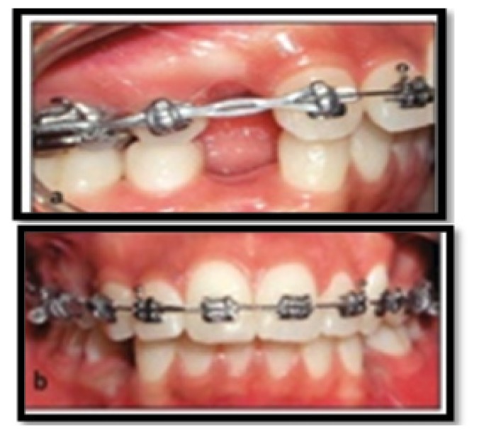
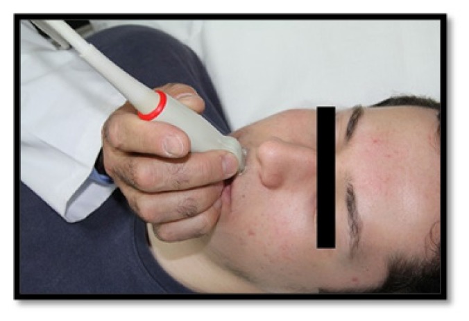
Background and aim: Orthodontic tooth movement (OTM) is a process whereby the application of a force induces bone resorption on the pressure side and bone apposition on the tension side of the lamina dura. However, only limited data are available on the in vivo behavior of the periodontal tissues. The aim of this study was to assess the changes of periodontal tissues, induced by the orthodontic canine retraction, using 40 MHz ultrasonography.
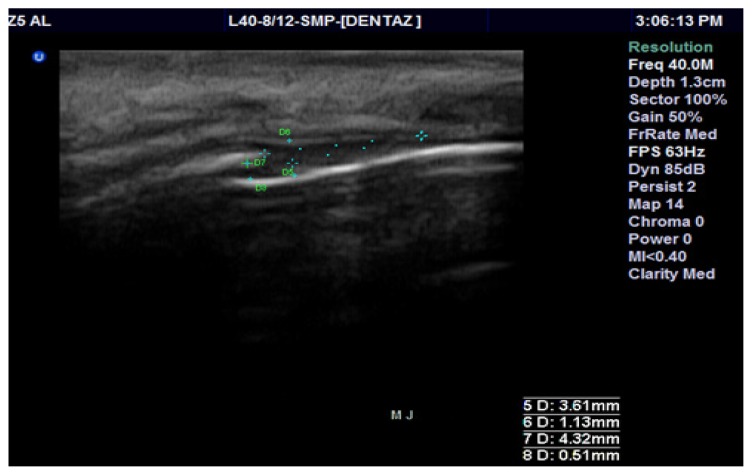
Methods: Ultrasonographic evaluation of periodontal tissues was conducted in 5 patients with indication for orthodontic treatment. The upper first premolars were extracted bilaterally due to severe crowding, and the canines were distalized using elastomeric chain with a net force of 100 cN. Ultrasonographic scans (US scans) were performed before, during and after retraction, in three distinct areas of the canines buccal surface: mesial, middle and distal. The reference point was the bracket, which appeared hyperechoic on the US scan. Four different dimensions were obtained: D1 (depth of the sulcus), D2 (thickness of the gingiva), D3 (length of the supracrestal fibers), D4 (width of periodontal space).
Results: An increase of D1 was observed in all three areas of the periodontium, during orthodontic treatment. D3 was strongly correlated before and immediately after force delivery only for the mesial area (r=0.828, p<0.05). In total, 228 variables were statistically analyzed using Pearson's correlation coefficients, in order to demonstrate the relationship between periodontal findings during orthodontic tooth movement.
Conclusion: High-resolution ultrasonography has the capability to obviate changes in periodontal ligament space and free gingiva during orthodontic tooth movement.

 求助内容:
求助内容: 应助结果提醒方式:


