Andrius Masedunskas, Mark Appaduray, Peter W Gunning, Edna C Hardeman
{"title":"点亮骨骼肌微管细胞骨架动力学。","authors":"Andrius Masedunskas, Mark Appaduray, Peter W Gunning, Edna C Hardeman","doi":"10.4161/intv.29293","DOIUrl":null,"url":null,"abstract":"<p><p>In the past few decades, live cell microscopy techniques in combination with fluorescent tagging have provided a true explosion in our knowledge of the inner functioning of the cell. Dynamic phenomena can be observed inside living cells and the behavior of individual molecules participating in those events can be documented. However, our preference for simple or easy model systems such as cell culture, has come at a cost of chasing artifacts and missing out on understanding real biology as it happens in complex multicellular organisms. We are now entering a new era where developing meaningful, but also tractable model systems to study biological phenomenon dynamically in vivo in a mammal is not only possible; it will become the gold standard for scientific quality and translational potential.<sup>1</sup><sup>,</sup><sup>2</sup> A study by Oddoux et al. describing the dynamics of the microtubule (MT) cytoskeleton in skeletal muscle is one example that demonstrates the power of developing in vivo/ex vivo models.<sup>3</sup> MTs have long attracted attention as targets for cancer therapeutics <sup>4</sup> and more recently as mediators of Duchene muscular dystrophy.<sup>5</sup> The muscle fiber MT cytoskeleton forms an intricate rectilinear lattice beneath the sarcolemma and is essential for the structural integrity of the muscle. Cultured cells do not develop such a specialized organization of the MT cytoskeleton and our understanding of it has come from static snapshots of muscle sections.<sup>6</sup> In this context, the methodology and the findings reported by Oddoux et al. are a significant step forward.</p>","PeriodicalId":14512,"journal":{"name":"IntraVital","volume":"3 1","pages":"e29293"},"PeriodicalIF":0.0000,"publicationDate":"2014-05-30","publicationTypes":"Journal Article","fieldsOfStudy":null,"isOpenAccess":false,"openAccessPdf":"https://sci-hub-pdf.com/10.4161/intv.29293","citationCount":"0","resultStr":"{\"title\":\"Lighting up microtubule cytoskeleton dynamics in skeletal muscle.\",\"authors\":\"Andrius Masedunskas, Mark Appaduray, Peter W Gunning, Edna C Hardeman\",\"doi\":\"10.4161/intv.29293\",\"DOIUrl\":null,\"url\":null,\"abstract\":\"<p><p>In the past few decades, live cell microscopy techniques in combination with fluorescent tagging have provided a true explosion in our knowledge of the inner functioning of the cell. Dynamic phenomena can be observed inside living cells and the behavior of individual molecules participating in those events can be documented. However, our preference for simple or easy model systems such as cell culture, has come at a cost of chasing artifacts and missing out on understanding real biology as it happens in complex multicellular organisms. We are now entering a new era where developing meaningful, but also tractable model systems to study biological phenomenon dynamically in vivo in a mammal is not only possible; it will become the gold standard for scientific quality and translational potential.<sup>1</sup><sup>,</sup><sup>2</sup> A study by Oddoux et al. describing the dynamics of the microtubule (MT) cytoskeleton in skeletal muscle is one example that demonstrates the power of developing in vivo/ex vivo models.<sup>3</sup> MTs have long attracted attention as targets for cancer therapeutics <sup>4</sup> and more recently as mediators of Duchene muscular dystrophy.<sup>5</sup> The muscle fiber MT cytoskeleton forms an intricate rectilinear lattice beneath the sarcolemma and is essential for the structural integrity of the muscle. Cultured cells do not develop such a specialized organization of the MT cytoskeleton and our understanding of it has come from static snapshots of muscle sections.<sup>6</sup> In this context, the methodology and the findings reported by Oddoux et al. are a significant step forward.</p>\",\"PeriodicalId\":14512,\"journal\":{\"name\":\"IntraVital\",\"volume\":\"3 1\",\"pages\":\"e29293\"},\"PeriodicalIF\":0.0000,\"publicationDate\":\"2014-05-30\",\"publicationTypes\":\"Journal Article\",\"fieldsOfStudy\":null,\"isOpenAccess\":false,\"openAccessPdf\":\"https://sci-hub-pdf.com/10.4161/intv.29293\",\"citationCount\":\"0\",\"resultStr\":null,\"platform\":\"Semanticscholar\",\"paperid\":null,\"PeriodicalName\":\"IntraVital\",\"FirstCategoryId\":\"1085\",\"ListUrlMain\":\"https://doi.org/10.4161/intv.29293\",\"RegionNum\":0,\"RegionCategory\":null,\"ArticlePicture\":[],\"TitleCN\":null,\"AbstractTextCN\":null,\"PMCID\":null,\"EPubDate\":\"2014/1/1 0:00:00\",\"PubModel\":\"eCollection\",\"JCR\":\"\",\"JCRName\":\"\",\"Score\":null,\"Total\":0}","platform":"Semanticscholar","paperid":null,"PeriodicalName":"IntraVital","FirstCategoryId":"1085","ListUrlMain":"https://doi.org/10.4161/intv.29293","RegionNum":0,"RegionCategory":null,"ArticlePicture":[],"TitleCN":null,"AbstractTextCN":null,"PMCID":null,"EPubDate":"2014/1/1 0:00:00","PubModel":"eCollection","JCR":"","JCRName":"","Score":null,"Total":0}
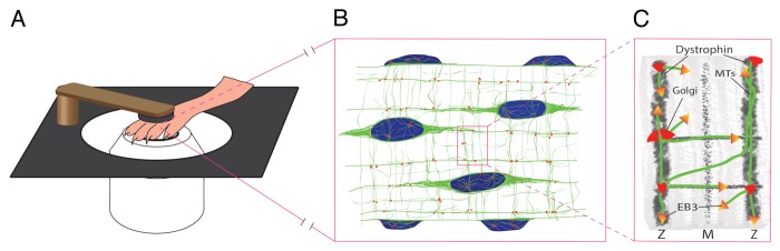
Lighting up microtubule cytoskeleton dynamics in skeletal muscle.
In the past few decades, live cell microscopy techniques in combination with fluorescent tagging have provided a true explosion in our knowledge of the inner functioning of the cell. Dynamic phenomena can be observed inside living cells and the behavior of individual molecules participating in those events can be documented. However, our preference for simple or easy model systems such as cell culture, has come at a cost of chasing artifacts and missing out on understanding real biology as it happens in complex multicellular organisms. We are now entering a new era where developing meaningful, but also tractable model systems to study biological phenomenon dynamically in vivo in a mammal is not only possible; it will become the gold standard for scientific quality and translational potential.1,2 A study by Oddoux et al. describing the dynamics of the microtubule (MT) cytoskeleton in skeletal muscle is one example that demonstrates the power of developing in vivo/ex vivo models.3 MTs have long attracted attention as targets for cancer therapeutics 4 and more recently as mediators of Duchene muscular dystrophy.5 The muscle fiber MT cytoskeleton forms an intricate rectilinear lattice beneath the sarcolemma and is essential for the structural integrity of the muscle. Cultured cells do not develop such a specialized organization of the MT cytoskeleton and our understanding of it has come from static snapshots of muscle sections.6 In this context, the methodology and the findings reported by Oddoux et al. are a significant step forward.

 求助内容:
求助内容: 应助结果提醒方式:


