{"title":"在脊髓小脑变性或多系统萎缩患者中,基于mri的小脑体积测量与国际合作性共济失调评定量表评分相关。","authors":"Daisuke Hara, Futaba Maki, Shigeaki Tanaka, Rie Sasaki, Yasuhiro Hasegawa","doi":"10.1186/s40673-016-0052-4","DOIUrl":null,"url":null,"abstract":"<p><strong>Background: </strong>Progression of clinical symptoms and cerebellar atrophy may vary among subtypes of spinocerebellar degeneration and multiple system atrophy. The aim of this cross-sectional study was to demonstrate the relationship between the International Cooperative Ataxia Rating Scale (ICARS) score and cerebellar volume derived from magnetic resonance imaging (MRI) in a broad spectrum of Japanese patients with cerebellar ataxia.</p><p><strong>Methods: </strong>A total of 86 patients with cerebellar ataxia (18 with cortical cerebellar atrophy, 34 with spinocerebellar ataxia, and 34 with multiple system atrophy) and 30 healthy subjects were studied. MRI-based cerebellar volume measurements were performed in all subjects using T1-weighted images acquired with a 1.5-T MRI scanner. The cerebellar volume/cranial anteroposterior (AP) diameter was used for statistical analysis.</p><p><strong>Results: </strong>Stepwise multiple regression analyses demonstrated that cerebellar volume/cranial AP diameter and midbrain AP/cranial AP diameter were significantly associated with the total score and domain I sub-score of ICARS. We found no interactions between these two anatomical factors in the ICARS total and domain I sub-scores. The main effects of these two predictors were statistically significant both in total and domain I sub-scores (p = 0.001 and 0.022, respectively).</p><p><strong>Conclusions: </strong>Cerebellar volume and midbrain AP diameter normalized to the cranial AP diameter were significantly correlated with the ICARS total and domain I sub-scores. Further longitudinal studies are warranted to explore the role of these MRI biomarkers for predicting disease progression.</p>","PeriodicalId":36752,"journal":{"name":"Cerebellum and Ataxias","volume":"3 ","pages":"14"},"PeriodicalIF":0.0000,"publicationDate":"2016-08-17","publicationTypes":"Journal Article","fieldsOfStudy":null,"isOpenAccess":false,"openAccessPdf":"https://sci-hub-pdf.com/10.1186/s40673-016-0052-4","citationCount":"15","resultStr":"{\"title\":\"MRI-based cerebellar volume measurements correlate with the International Cooperative Ataxia Rating Scale score in patients with spinocerebellar degeneration or multiple system atrophy.\",\"authors\":\"Daisuke Hara, Futaba Maki, Shigeaki Tanaka, Rie Sasaki, Yasuhiro Hasegawa\",\"doi\":\"10.1186/s40673-016-0052-4\",\"DOIUrl\":null,\"url\":null,\"abstract\":\"<p><strong>Background: </strong>Progression of clinical symptoms and cerebellar atrophy may vary among subtypes of spinocerebellar degeneration and multiple system atrophy. The aim of this cross-sectional study was to demonstrate the relationship between the International Cooperative Ataxia Rating Scale (ICARS) score and cerebellar volume derived from magnetic resonance imaging (MRI) in a broad spectrum of Japanese patients with cerebellar ataxia.</p><p><strong>Methods: </strong>A total of 86 patients with cerebellar ataxia (18 with cortical cerebellar atrophy, 34 with spinocerebellar ataxia, and 34 with multiple system atrophy) and 30 healthy subjects were studied. MRI-based cerebellar volume measurements were performed in all subjects using T1-weighted images acquired with a 1.5-T MRI scanner. The cerebellar volume/cranial anteroposterior (AP) diameter was used for statistical analysis.</p><p><strong>Results: </strong>Stepwise multiple regression analyses demonstrated that cerebellar volume/cranial AP diameter and midbrain AP/cranial AP diameter were significantly associated with the total score and domain I sub-score of ICARS. We found no interactions between these two anatomical factors in the ICARS total and domain I sub-scores. The main effects of these two predictors were statistically significant both in total and domain I sub-scores (p = 0.001 and 0.022, respectively).</p><p><strong>Conclusions: </strong>Cerebellar volume and midbrain AP diameter normalized to the cranial AP diameter were significantly correlated with the ICARS total and domain I sub-scores. Further longitudinal studies are warranted to explore the role of these MRI biomarkers for predicting disease progression.</p>\",\"PeriodicalId\":36752,\"journal\":{\"name\":\"Cerebellum and Ataxias\",\"volume\":\"3 \",\"pages\":\"14\"},\"PeriodicalIF\":0.0000,\"publicationDate\":\"2016-08-17\",\"publicationTypes\":\"Journal Article\",\"fieldsOfStudy\":null,\"isOpenAccess\":false,\"openAccessPdf\":\"https://sci-hub-pdf.com/10.1186/s40673-016-0052-4\",\"citationCount\":\"15\",\"resultStr\":null,\"platform\":\"Semanticscholar\",\"paperid\":null,\"PeriodicalName\":\"Cerebellum and Ataxias\",\"FirstCategoryId\":\"1085\",\"ListUrlMain\":\"https://doi.org/10.1186/s40673-016-0052-4\",\"RegionNum\":0,\"RegionCategory\":null,\"ArticlePicture\":[],\"TitleCN\":null,\"AbstractTextCN\":null,\"PMCID\":null,\"EPubDate\":\"2016/1/1 0:00:00\",\"PubModel\":\"eCollection\",\"JCR\":\"Q3\",\"JCRName\":\"Medicine\",\"Score\":null,\"Total\":0}","platform":"Semanticscholar","paperid":null,"PeriodicalName":"Cerebellum and Ataxias","FirstCategoryId":"1085","ListUrlMain":"https://doi.org/10.1186/s40673-016-0052-4","RegionNum":0,"RegionCategory":null,"ArticlePicture":[],"TitleCN":null,"AbstractTextCN":null,"PMCID":null,"EPubDate":"2016/1/1 0:00:00","PubModel":"eCollection","JCR":"Q3","JCRName":"Medicine","Score":null,"Total":0}
引用次数: 15
摘要
背景:脊髓小脑变性和多系统萎缩不同亚型的临床症状和小脑萎缩的进展可能不同。本横断面研究的目的是证明国际合作共济失调评定量表(ICARS)评分与日本小脑共济失调患者的广谱磁共振成像(MRI)小脑体积之间的关系。方法:选取86例小脑共济失调患者(皮质性小脑萎缩18例、脊髓性小脑共济失调34例、多系统萎缩34例)和30例健康对照。所有受试者使用1.5 t MRI扫描仪获得的t1加权图像进行基于MRI的小脑体积测量。采用小脑体积/颅前径进行统计学分析。结果:逐步多元回归分析显示,小脑体积/颅AP直径和中脑AP/颅AP直径与ICARS总分和I区分有显著相关。我们发现这两个解剖因素在ICARS总分和域I分评分中没有相互作用。这两个预测因子的主效应在总分和域1分上均有统计学意义(p分别= 0.001和0.022)。结论:小脑体积和中脑AP直径与ICARS总分和I区分有显著相关。进一步的纵向研究有必要探索这些MRI生物标志物在预测疾病进展方面的作用。
MRI-based cerebellar volume measurements correlate with the International Cooperative Ataxia Rating Scale score in patients with spinocerebellar degeneration or multiple system atrophy.
Background: Progression of clinical symptoms and cerebellar atrophy may vary among subtypes of spinocerebellar degeneration and multiple system atrophy. The aim of this cross-sectional study was to demonstrate the relationship between the International Cooperative Ataxia Rating Scale (ICARS) score and cerebellar volume derived from magnetic resonance imaging (MRI) in a broad spectrum of Japanese patients with cerebellar ataxia.
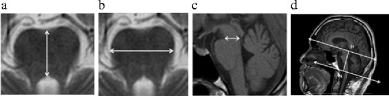
Methods: A total of 86 patients with cerebellar ataxia (18 with cortical cerebellar atrophy, 34 with spinocerebellar ataxia, and 34 with multiple system atrophy) and 30 healthy subjects were studied. MRI-based cerebellar volume measurements were performed in all subjects using T1-weighted images acquired with a 1.5-T MRI scanner. The cerebellar volume/cranial anteroposterior (AP) diameter was used for statistical analysis.
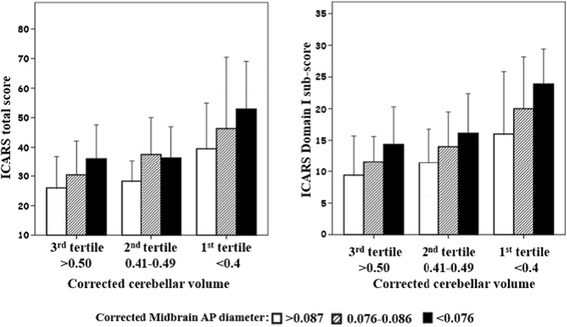
Results: Stepwise multiple regression analyses demonstrated that cerebellar volume/cranial AP diameter and midbrain AP/cranial AP diameter were significantly associated with the total score and domain I sub-score of ICARS. We found no interactions between these two anatomical factors in the ICARS total and domain I sub-scores. The main effects of these two predictors were statistically significant both in total and domain I sub-scores (p = 0.001 and 0.022, respectively).
Conclusions: Cerebellar volume and midbrain AP diameter normalized to the cranial AP diameter were significantly correlated with the ICARS total and domain I sub-scores. Further longitudinal studies are warranted to explore the role of these MRI biomarkers for predicting disease progression.


 求助内容:
求助内容: 应助结果提醒方式:


