Christian L Roth, Gabrielle D'Ambrosio, Clinton Elfers
{"title":"下丘脑损伤后核因子κ B通路的激活和下丘脑催产素的减少。","authors":"Christian L Roth, Gabrielle D'Ambrosio, Clinton Elfers","doi":"10.15761/JSIN.1000114","DOIUrl":null,"url":null,"abstract":"<p><strong>Background: </strong>Hypothalamic obesity (HO) occurs in patients with tumors and lesions in the medial hypothalamic region. In this study, a hyperphagic rat model of combined medial hypothalamic lesions (CMHL) was used to test which specific inflammatory molecules are involved.</p><p><strong>Methods: </strong>In order to target specific homeostatic medial hypothalamic nuclei (arcuate, ventromedial, and dorsomedial nuclei), male Sprague-Dawley rats (age of 8 weeks, ~250 g body weight) received four electrolytic lesions or sham surgery. Post-surgery food intake and weight changes were tracked and hypothalamic gene expression for inflammatory molecules as well as anorexigenic peptide oxytocin 7 days and 7 months post-surgery were tested.</p><p><strong>Results: </strong>Seven days post-surgery, average food intake increased by 23%, and body weight gain had increased by 68%. Toll-like 4 receptor/nuclear factor-κB (TLR4/NF-κB)-pathway was specifically activated in the mediobasal hypothalamus (MBH), resulting in 3-fold higher tumor necrosis factor (TNF)-α, 10-fold higher interleukin (IL) 1-β mRNA levels, and higher expression of suppression of cytokine signaling (SOCS) 3, while oxytocin mRNA levels were significantly reduced in CMHL rats versus sham surgery rats 7 days post-surgery. At 7 months, inflammation was less stimulated in MBH of CMHL rats compared to 7 days post-surgery and SOCS 3 as well as oxytocin mRNA levels were comparable between the two groups.</p><p><strong>Conclusion: </strong>Medial hypothalamic lesions are associated with strong post-surgery hyperphagia and activation of TLR4/NF-κB-pathway as well as reduced expression of oxytocin in the hypothalamus.</p>","PeriodicalId":87318,"journal":{"name":"Journal of systems and integrative neuroscience","volume":"2 1","pages":"79-84"},"PeriodicalIF":0.0000,"publicationDate":"2016-02-01","publicationTypes":"Journal Article","fieldsOfStudy":null,"isOpenAccess":false,"openAccessPdf":"https://www.ncbi.nlm.nih.gov/pmc/articles/PMC4976786/pdf/","citationCount":"7","resultStr":"{\"title\":\"Activation of nuclear factor kappa B pathway and reduction of hypothalamic oxytocin following hypothalamic lesions.\",\"authors\":\"Christian L Roth, Gabrielle D'Ambrosio, Clinton Elfers\",\"doi\":\"10.15761/JSIN.1000114\",\"DOIUrl\":null,\"url\":null,\"abstract\":\"<p><strong>Background: </strong>Hypothalamic obesity (HO) occurs in patients with tumors and lesions in the medial hypothalamic region. In this study, a hyperphagic rat model of combined medial hypothalamic lesions (CMHL) was used to test which specific inflammatory molecules are involved.</p><p><strong>Methods: </strong>In order to target specific homeostatic medial hypothalamic nuclei (arcuate, ventromedial, and dorsomedial nuclei), male Sprague-Dawley rats (age of 8 weeks, ~250 g body weight) received four electrolytic lesions or sham surgery. Post-surgery food intake and weight changes were tracked and hypothalamic gene expression for inflammatory molecules as well as anorexigenic peptide oxytocin 7 days and 7 months post-surgery were tested.</p><p><strong>Results: </strong>Seven days post-surgery, average food intake increased by 23%, and body weight gain had increased by 68%. Toll-like 4 receptor/nuclear factor-κB (TLR4/NF-κB)-pathway was specifically activated in the mediobasal hypothalamus (MBH), resulting in 3-fold higher tumor necrosis factor (TNF)-α, 10-fold higher interleukin (IL) 1-β mRNA levels, and higher expression of suppression of cytokine signaling (SOCS) 3, while oxytocin mRNA levels were significantly reduced in CMHL rats versus sham surgery rats 7 days post-surgery. At 7 months, inflammation was less stimulated in MBH of CMHL rats compared to 7 days post-surgery and SOCS 3 as well as oxytocin mRNA levels were comparable between the two groups.</p><p><strong>Conclusion: </strong>Medial hypothalamic lesions are associated with strong post-surgery hyperphagia and activation of TLR4/NF-κB-pathway as well as reduced expression of oxytocin in the hypothalamus.</p>\",\"PeriodicalId\":87318,\"journal\":{\"name\":\"Journal of systems and integrative neuroscience\",\"volume\":\"2 1\",\"pages\":\"79-84\"},\"PeriodicalIF\":0.0000,\"publicationDate\":\"2016-02-01\",\"publicationTypes\":\"Journal Article\",\"fieldsOfStudy\":null,\"isOpenAccess\":false,\"openAccessPdf\":\"https://www.ncbi.nlm.nih.gov/pmc/articles/PMC4976786/pdf/\",\"citationCount\":\"7\",\"resultStr\":null,\"platform\":\"Semanticscholar\",\"paperid\":null,\"PeriodicalName\":\"Journal of systems and integrative neuroscience\",\"FirstCategoryId\":\"1085\",\"ListUrlMain\":\"https://doi.org/10.15761/JSIN.1000114\",\"RegionNum\":0,\"RegionCategory\":null,\"ArticlePicture\":[],\"TitleCN\":null,\"AbstractTextCN\":null,\"PMCID\":null,\"EPubDate\":\"2016/1/29 0:00:00\",\"PubModel\":\"Epub\",\"JCR\":\"\",\"JCRName\":\"\",\"Score\":null,\"Total\":0}","platform":"Semanticscholar","paperid":null,"PeriodicalName":"Journal of systems and integrative neuroscience","FirstCategoryId":"1085","ListUrlMain":"https://doi.org/10.15761/JSIN.1000114","RegionNum":0,"RegionCategory":null,"ArticlePicture":[],"TitleCN":null,"AbstractTextCN":null,"PMCID":null,"EPubDate":"2016/1/29 0:00:00","PubModel":"Epub","JCR":"","JCRName":"","Score":null,"Total":0}
Activation of nuclear factor kappa B pathway and reduction of hypothalamic oxytocin following hypothalamic lesions.
Background: Hypothalamic obesity (HO) occurs in patients with tumors and lesions in the medial hypothalamic region. In this study, a hyperphagic rat model of combined medial hypothalamic lesions (CMHL) was used to test which specific inflammatory molecules are involved.
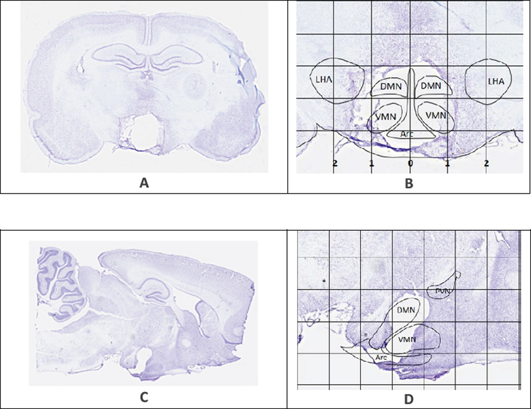
Methods: In order to target specific homeostatic medial hypothalamic nuclei (arcuate, ventromedial, and dorsomedial nuclei), male Sprague-Dawley rats (age of 8 weeks, ~250 g body weight) received four electrolytic lesions or sham surgery. Post-surgery food intake and weight changes were tracked and hypothalamic gene expression for inflammatory molecules as well as anorexigenic peptide oxytocin 7 days and 7 months post-surgery were tested.
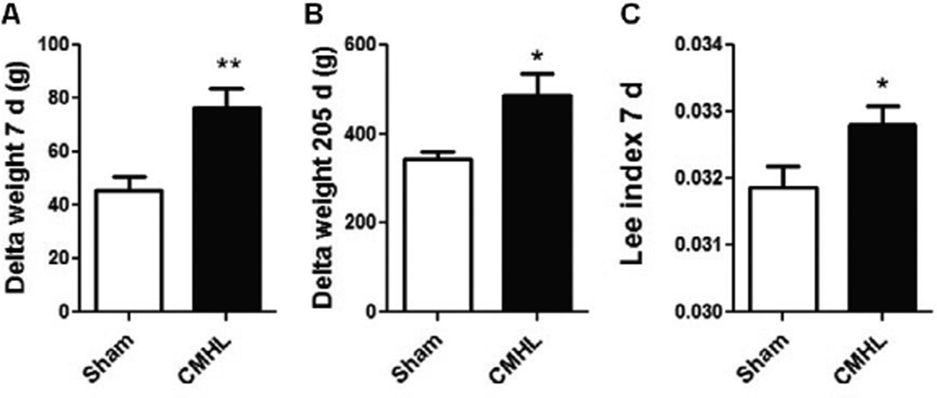
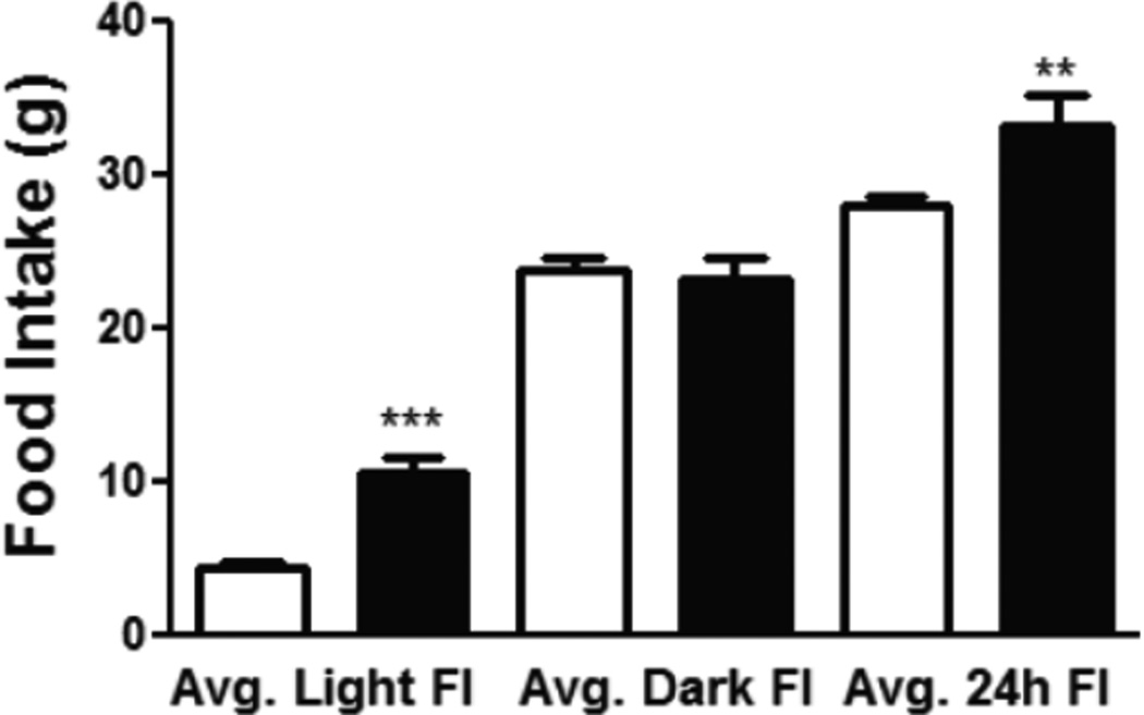
Results: Seven days post-surgery, average food intake increased by 23%, and body weight gain had increased by 68%. Toll-like 4 receptor/nuclear factor-κB (TLR4/NF-κB)-pathway was specifically activated in the mediobasal hypothalamus (MBH), resulting in 3-fold higher tumor necrosis factor (TNF)-α, 10-fold higher interleukin (IL) 1-β mRNA levels, and higher expression of suppression of cytokine signaling (SOCS) 3, while oxytocin mRNA levels were significantly reduced in CMHL rats versus sham surgery rats 7 days post-surgery. At 7 months, inflammation was less stimulated in MBH of CMHL rats compared to 7 days post-surgery and SOCS 3 as well as oxytocin mRNA levels were comparable between the two groups.
Conclusion: Medial hypothalamic lesions are associated with strong post-surgery hyperphagia and activation of TLR4/NF-κB-pathway as well as reduced expression of oxytocin in the hypothalamus.

 求助内容:
求助内容: 应助结果提醒方式:


