Aida Sabaté-Llobera, Montserrat Cortés-Romera, Santiago Mercadal, Javier Hernández-Gañán, Helena Pomares, Eva González-Barca, Cristina Gámez-Cenzano
{"title":"局部弥漫性大b细胞淋巴瘤初始分期的低剂量PET/CT和全剂量增强CT。","authors":"Aida Sabaté-Llobera, Montserrat Cortés-Romera, Santiago Mercadal, Javier Hernández-Gañán, Helena Pomares, Eva González-Barca, Cristina Gámez-Cenzano","doi":"10.4137/CMBD.S38468","DOIUrl":null,"url":null,"abstract":"<p><p>Computed tomography (CT) has been used as the reference imaging technique for the initial staging of diffuse large B-cell lymphoma until recent days, when the introduction of positron emission tomography (PET)/CT imaging as a hybrid technique has become of routine use. However, the performance of both examinations is still common. The aim of this work was to compare the findings between low-dose 2-deoxy-2-((18)F)fluoro-d-glucose ((18)F-FDG) PET/CT and full-dose contrast-enhanced CT (ceCT) in 28 patients with localized diffuse large B-cell lymphoma according to PET/CT findings, in order to avoid the performance of ceCT. For each technique, a comparison in the number of nodal and extranodal involved regions was performed. PET/CT showed more lesions than ceCT in both nodal (41 vs. 36) and extranodal localizations (16 vs. 15). Disease staging according to both techniques was concordant in 22 patients (79%) and discordant in 6 patients (21%), changing treatment management in 3 patients (11%). PET/CT determined a better staging and therapeutic approach, making the performance of an additional ceCT unnecessary. </p>","PeriodicalId":43083,"journal":{"name":"Clinical Medicine Insights-Blood Disorders","volume":"9 ","pages":"29-32"},"PeriodicalIF":3.0000,"publicationDate":"2016-08-17","publicationTypes":"Journal Article","fieldsOfStudy":null,"isOpenAccess":false,"openAccessPdf":"https://sci-hub-pdf.com/10.4137/CMBD.S38468","citationCount":"7","resultStr":"{\"title\":\"Low-Dose PET/CT and Full-Dose Contrast-Enhanced CT at the Initial Staging of Localized Diffuse Large B-Cell Lymphomas.\",\"authors\":\"Aida Sabaté-Llobera, Montserrat Cortés-Romera, Santiago Mercadal, Javier Hernández-Gañán, Helena Pomares, Eva González-Barca, Cristina Gámez-Cenzano\",\"doi\":\"10.4137/CMBD.S38468\",\"DOIUrl\":null,\"url\":null,\"abstract\":\"<p><p>Computed tomography (CT) has been used as the reference imaging technique for the initial staging of diffuse large B-cell lymphoma until recent days, when the introduction of positron emission tomography (PET)/CT imaging as a hybrid technique has become of routine use. However, the performance of both examinations is still common. The aim of this work was to compare the findings between low-dose 2-deoxy-2-((18)F)fluoro-d-glucose ((18)F-FDG) PET/CT and full-dose contrast-enhanced CT (ceCT) in 28 patients with localized diffuse large B-cell lymphoma according to PET/CT findings, in order to avoid the performance of ceCT. For each technique, a comparison in the number of nodal and extranodal involved regions was performed. PET/CT showed more lesions than ceCT in both nodal (41 vs. 36) and extranodal localizations (16 vs. 15). Disease staging according to both techniques was concordant in 22 patients (79%) and discordant in 6 patients (21%), changing treatment management in 3 patients (11%). PET/CT determined a better staging and therapeutic approach, making the performance of an additional ceCT unnecessary. </p>\",\"PeriodicalId\":43083,\"journal\":{\"name\":\"Clinical Medicine Insights-Blood Disorders\",\"volume\":\"9 \",\"pages\":\"29-32\"},\"PeriodicalIF\":3.0000,\"publicationDate\":\"2016-08-17\",\"publicationTypes\":\"Journal Article\",\"fieldsOfStudy\":null,\"isOpenAccess\":false,\"openAccessPdf\":\"https://sci-hub-pdf.com/10.4137/CMBD.S38468\",\"citationCount\":\"7\",\"resultStr\":null,\"platform\":\"Semanticscholar\",\"paperid\":null,\"PeriodicalName\":\"Clinical Medicine Insights-Blood Disorders\",\"FirstCategoryId\":\"1085\",\"ListUrlMain\":\"https://doi.org/10.4137/CMBD.S38468\",\"RegionNum\":0,\"RegionCategory\":null,\"ArticlePicture\":[],\"TitleCN\":null,\"AbstractTextCN\":null,\"PMCID\":null,\"EPubDate\":\"2016/1/1 0:00:00\",\"PubModel\":\"eCollection\",\"JCR\":\"Q2\",\"JCRName\":\"Medicine\",\"Score\":null,\"Total\":0}","platform":"Semanticscholar","paperid":null,"PeriodicalName":"Clinical Medicine Insights-Blood Disorders","FirstCategoryId":"1085","ListUrlMain":"https://doi.org/10.4137/CMBD.S38468","RegionNum":0,"RegionCategory":null,"ArticlePicture":[],"TitleCN":null,"AbstractTextCN":null,"PMCID":null,"EPubDate":"2016/1/1 0:00:00","PubModel":"eCollection","JCR":"Q2","JCRName":"Medicine","Score":null,"Total":0}
Low-Dose PET/CT and Full-Dose Contrast-Enhanced CT at the Initial Staging of Localized Diffuse Large B-Cell Lymphomas.
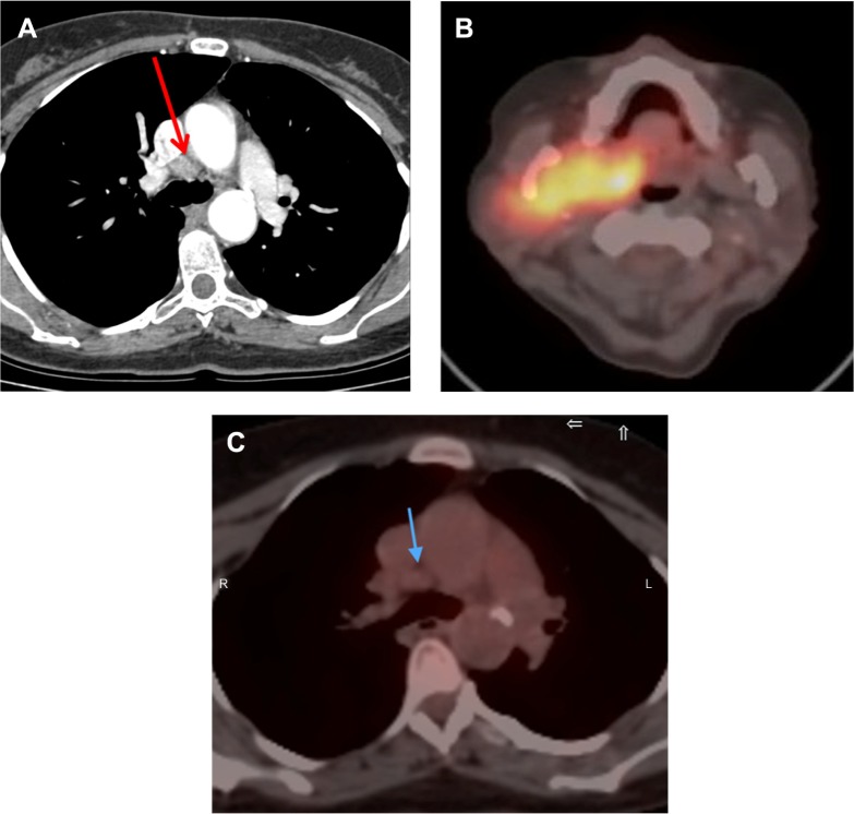
Computed tomography (CT) has been used as the reference imaging technique for the initial staging of diffuse large B-cell lymphoma until recent days, when the introduction of positron emission tomography (PET)/CT imaging as a hybrid technique has become of routine use. However, the performance of both examinations is still common. The aim of this work was to compare the findings between low-dose 2-deoxy-2-((18)F)fluoro-d-glucose ((18)F-FDG) PET/CT and full-dose contrast-enhanced CT (ceCT) in 28 patients with localized diffuse large B-cell lymphoma according to PET/CT findings, in order to avoid the performance of ceCT. For each technique, a comparison in the number of nodal and extranodal involved regions was performed. PET/CT showed more lesions than ceCT in both nodal (41 vs. 36) and extranodal localizations (16 vs. 15). Disease staging according to both techniques was concordant in 22 patients (79%) and discordant in 6 patients (21%), changing treatment management in 3 patients (11%). PET/CT determined a better staging and therapeutic approach, making the performance of an additional ceCT unnecessary.

 求助内容:
求助内容: 应助结果提醒方式:


