Rebecca J Crawford, Thomas Volken, Stephanie Valentin, Markus Melloh, James M Elliott
{"title":"无症状人群腰椎椎旁肌脂肪浸润率与脊柱退变:一项年龄聚集的横断面模拟研究。","authors":"Rebecca J Crawford, Thomas Volken, Stephanie Valentin, Markus Melloh, James M Elliott","doi":"10.1186/s13013-016-0080-0","DOIUrl":null,"url":null,"abstract":"<p><strong>Background: </strong>The spinal column including its vertebrae and disks has been well examined and extensively reported in relation to age-aggregated degeneration. In contrast, paravertebral muscles are poorly represented in describing normative degeneration. Increasing evidence points to the importance of paravertebral muscle quality in low back health, and their potential as a modifiable factor in low back pain (LBP). Studies examining normative decline of paravertebral muscles are needed to advance the field's etiological understanding. With a novel approach and based on published data, we establish and compare decline rates of imaging features for degeneration of lumbar vertebrae and disks, versus fatty infiltration in paravertebral muscles in asymptomatic adults.</p><p><strong>Methods: </strong>Our cross-sectional simulation study examined age-aggregated data from three published studies who reported on asymptomatic adults spanning 18-60 years. Prevalence rates of imaging degenerative features of the spinal column were examined via logistic regression and compared with percentage fatty infiltration in erector spinae, multifidus and psoas using synthetic data and Monte Carlo simulation with 10,000 endpoint-specific regression iterations. General linear regression models were employed to estimate marginal effects of age reported as a one-year change rate (with 95 % confidence intervals) for comparisons between all reported spinal features.</p><p><strong>Results: </strong>Declines in multifidus (0.24 & 0.11 %/year), erector spinae (0.13 & 0.07 %/year), and psoas (0.04 %/year) occur at similarly slow rates to disk protrusion (0.25 %/year), annular fissure (0.15 %/year), and spondylolisthesis (0.29 %/year). Multifidus showed a trend for faster decline than erector spinae, particularly in men. Of the features examined, disk signal loss declined fastest, and psoas muscle the slowest.</p><p><strong>Conclusions: </strong>Degeneration of lumbar paravertebral muscles occurs slowly in asymptomatic adults, with a tendency to be most pronounced in multifidus. Rate of decline of spinal structures represents a novel variable that warrants inclusion as a known feature of the expected degenerative cascade, and to provide a basis for comparison to diseases of the spine in research and clinical practice. Concurrent examination of spinal features using advanced imaging to improve muscle analysis would be a strong addition to the field.</p>","PeriodicalId":21573,"journal":{"name":"Scoliosis and Spinal Disorders","volume":"11 ","pages":"21"},"PeriodicalIF":0.0000,"publicationDate":"2016-08-05","publicationTypes":"Journal Article","fieldsOfStudy":null,"isOpenAccess":false,"openAccessPdf":"https://sci-hub-pdf.com/10.1186/s13013-016-0080-0","citationCount":"43","resultStr":"{\"title\":\"Rate of lumbar paravertebral muscle fat infiltration versus spinal degeneration in asymptomatic populations: an age-aggregated cross-sectional simulation study.\",\"authors\":\"Rebecca J Crawford, Thomas Volken, Stephanie Valentin, Markus Melloh, James M Elliott\",\"doi\":\"10.1186/s13013-016-0080-0\",\"DOIUrl\":null,\"url\":null,\"abstract\":\"<p><strong>Background: </strong>The spinal column including its vertebrae and disks has been well examined and extensively reported in relation to age-aggregated degeneration. In contrast, paravertebral muscles are poorly represented in describing normative degeneration. Increasing evidence points to the importance of paravertebral muscle quality in low back health, and their potential as a modifiable factor in low back pain (LBP). Studies examining normative decline of paravertebral muscles are needed to advance the field's etiological understanding. With a novel approach and based on published data, we establish and compare decline rates of imaging features for degeneration of lumbar vertebrae and disks, versus fatty infiltration in paravertebral muscles in asymptomatic adults.</p><p><strong>Methods: </strong>Our cross-sectional simulation study examined age-aggregated data from three published studies who reported on asymptomatic adults spanning 18-60 years. Prevalence rates of imaging degenerative features of the spinal column were examined via logistic regression and compared with percentage fatty infiltration in erector spinae, multifidus and psoas using synthetic data and Monte Carlo simulation with 10,000 endpoint-specific regression iterations. General linear regression models were employed to estimate marginal effects of age reported as a one-year change rate (with 95 % confidence intervals) for comparisons between all reported spinal features.</p><p><strong>Results: </strong>Declines in multifidus (0.24 & 0.11 %/year), erector spinae (0.13 & 0.07 %/year), and psoas (0.04 %/year) occur at similarly slow rates to disk protrusion (0.25 %/year), annular fissure (0.15 %/year), and spondylolisthesis (0.29 %/year). Multifidus showed a trend for faster decline than erector spinae, particularly in men. Of the features examined, disk signal loss declined fastest, and psoas muscle the slowest.</p><p><strong>Conclusions: </strong>Degeneration of lumbar paravertebral muscles occurs slowly in asymptomatic adults, with a tendency to be most pronounced in multifidus. Rate of decline of spinal structures represents a novel variable that warrants inclusion as a known feature of the expected degenerative cascade, and to provide a basis for comparison to diseases of the spine in research and clinical practice. Concurrent examination of spinal features using advanced imaging to improve muscle analysis would be a strong addition to the field.</p>\",\"PeriodicalId\":21573,\"journal\":{\"name\":\"Scoliosis and Spinal Disorders\",\"volume\":\"11 \",\"pages\":\"21\"},\"PeriodicalIF\":0.0000,\"publicationDate\":\"2016-08-05\",\"publicationTypes\":\"Journal Article\",\"fieldsOfStudy\":null,\"isOpenAccess\":false,\"openAccessPdf\":\"https://sci-hub-pdf.com/10.1186/s13013-016-0080-0\",\"citationCount\":\"43\",\"resultStr\":null,\"platform\":\"Semanticscholar\",\"paperid\":null,\"PeriodicalName\":\"Scoliosis and Spinal Disorders\",\"FirstCategoryId\":\"1085\",\"ListUrlMain\":\"https://doi.org/10.1186/s13013-016-0080-0\",\"RegionNum\":0,\"RegionCategory\":null,\"ArticlePicture\":[],\"TitleCN\":null,\"AbstractTextCN\":null,\"PMCID\":null,\"EPubDate\":\"2016/1/1 0:00:00\",\"PubModel\":\"eCollection\",\"JCR\":\"Q1\",\"JCRName\":\"Medicine\",\"Score\":null,\"Total\":0}","platform":"Semanticscholar","paperid":null,"PeriodicalName":"Scoliosis and Spinal Disorders","FirstCategoryId":"1085","ListUrlMain":"https://doi.org/10.1186/s13013-016-0080-0","RegionNum":0,"RegionCategory":null,"ArticlePicture":[],"TitleCN":null,"AbstractTextCN":null,"PMCID":null,"EPubDate":"2016/1/1 0:00:00","PubModel":"eCollection","JCR":"Q1","JCRName":"Medicine","Score":null,"Total":0}
Rate of lumbar paravertebral muscle fat infiltration versus spinal degeneration in asymptomatic populations: an age-aggregated cross-sectional simulation study.
Background: The spinal column including its vertebrae and disks has been well examined and extensively reported in relation to age-aggregated degeneration. In contrast, paravertebral muscles are poorly represented in describing normative degeneration. Increasing evidence points to the importance of paravertebral muscle quality in low back health, and their potential as a modifiable factor in low back pain (LBP). Studies examining normative decline of paravertebral muscles are needed to advance the field's etiological understanding. With a novel approach and based on published data, we establish and compare decline rates of imaging features for degeneration of lumbar vertebrae and disks, versus fatty infiltration in paravertebral muscles in asymptomatic adults.
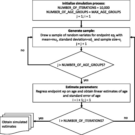
Methods: Our cross-sectional simulation study examined age-aggregated data from three published studies who reported on asymptomatic adults spanning 18-60 years. Prevalence rates of imaging degenerative features of the spinal column were examined via logistic regression and compared with percentage fatty infiltration in erector spinae, multifidus and psoas using synthetic data and Monte Carlo simulation with 10,000 endpoint-specific regression iterations. General linear regression models were employed to estimate marginal effects of age reported as a one-year change rate (with 95 % confidence intervals) for comparisons between all reported spinal features.
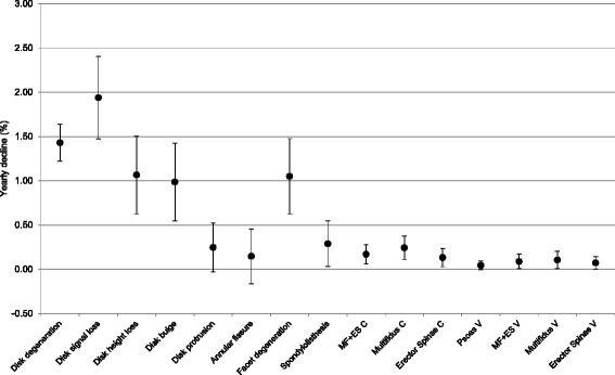
Results: Declines in multifidus (0.24 & 0.11 %/year), erector spinae (0.13 & 0.07 %/year), and psoas (0.04 %/year) occur at similarly slow rates to disk protrusion (0.25 %/year), annular fissure (0.15 %/year), and spondylolisthesis (0.29 %/year). Multifidus showed a trend for faster decline than erector spinae, particularly in men. Of the features examined, disk signal loss declined fastest, and psoas muscle the slowest.
Conclusions: Degeneration of lumbar paravertebral muscles occurs slowly in asymptomatic adults, with a tendency to be most pronounced in multifidus. Rate of decline of spinal structures represents a novel variable that warrants inclusion as a known feature of the expected degenerative cascade, and to provide a basis for comparison to diseases of the spine in research and clinical practice. Concurrent examination of spinal features using advanced imaging to improve muscle analysis would be a strong addition to the field.
期刊介绍:
Cessation.Scoliosis and Spinal Disorders is an open access, multidisciplinary journal that encompasses all aspects of research on prevention, diagnosis, treatment, outcomes and cost-analyses of conservative and surgical management of all spinal deformities and disorders. Both clinical and basic science reports form the cornerstone of the journal in its endeavour to provide original, primary studies as well as narrative/systematic reviews and meta-analyses to the academic community and beyond. Scoliosis and Spinal Disorders aims to provide an integrated and balanced view of cutting-edge spine research to further enhance effective collaboration among clinical spine specialists and scientists, and to ultimately improve patient outcomes based on an evidence-based spine care approach.

 求助内容:
求助内容: 应助结果提醒方式:


