Doo-Ho Lee, Young Joon Ahn, Rumi Shin, Hae Won Lee
{"title":"胆管远端转移性粘液腺癌,来自横断结肠癌,表现为梗阻性黄疸。","authors":"Doo-Ho Lee, Young Joon Ahn, Rumi Shin, Hae Won Lee","doi":"10.14701/kjhbps.2015.19.3.125","DOIUrl":null,"url":null,"abstract":"<p><p>The patient was a 70-year-old male whose chief complaints were obstructive jaundice and weight loss. Abdominal imaging studies showed a 2.5 cm sized mass at the distal common bile duct, which was suggestive of bile duct cancer. Eccentric enhancing wall thickening in the transverse colon was also shown, suggesting concomitant colon cancer. A colonoscopy revealed a lumen-encircling ulcerofungating mass in the transverse colon, that was pathologically proven to be adenocarcinoma. The bile duct pathology was also adenocarcinoma. Pylorus-preserving pancreaticoduodenectomy and extended right hemicolectomy were performed under the diagnosis of double primary cancers. Postoperative histopathologic examination revealed moderately differentiated mucinous adenocarcinoma of transverse colon cancer, and mucinous adenocarcinoma of the distal common bile duct. Immunohistochemical staining studies showed that the bile duct cancer had metastasized from the colon cancer. The patient recovered uneventfully from surgery and will be undergoing chemotherapy for three months. </p>","PeriodicalId":91136,"journal":{"name":"Korean journal of hepato-biliary-pancreatic surgery","volume":"19 3","pages":"125-8"},"PeriodicalIF":0.0000,"publicationDate":"2015-08-01","publicationTypes":"Journal Article","fieldsOfStudy":null,"isOpenAccess":false,"openAccessPdf":"https://sci-hub-pdf.com/10.14701/kjhbps.2015.19.3.125","citationCount":"7","resultStr":"{\"title\":\"Metastatic mucinous adenocarcinoma of the distal common bile duct, from transverse colon cancer presenting as obstructive jaundice.\",\"authors\":\"Doo-Ho Lee, Young Joon Ahn, Rumi Shin, Hae Won Lee\",\"doi\":\"10.14701/kjhbps.2015.19.3.125\",\"DOIUrl\":null,\"url\":null,\"abstract\":\"<p><p>The patient was a 70-year-old male whose chief complaints were obstructive jaundice and weight loss. Abdominal imaging studies showed a 2.5 cm sized mass at the distal common bile duct, which was suggestive of bile duct cancer. Eccentric enhancing wall thickening in the transverse colon was also shown, suggesting concomitant colon cancer. A colonoscopy revealed a lumen-encircling ulcerofungating mass in the transverse colon, that was pathologically proven to be adenocarcinoma. The bile duct pathology was also adenocarcinoma. Pylorus-preserving pancreaticoduodenectomy and extended right hemicolectomy were performed under the diagnosis of double primary cancers. Postoperative histopathologic examination revealed moderately differentiated mucinous adenocarcinoma of transverse colon cancer, and mucinous adenocarcinoma of the distal common bile duct. Immunohistochemical staining studies showed that the bile duct cancer had metastasized from the colon cancer. The patient recovered uneventfully from surgery and will be undergoing chemotherapy for three months. </p>\",\"PeriodicalId\":91136,\"journal\":{\"name\":\"Korean journal of hepato-biliary-pancreatic surgery\",\"volume\":\"19 3\",\"pages\":\"125-8\"},\"PeriodicalIF\":0.0000,\"publicationDate\":\"2015-08-01\",\"publicationTypes\":\"Journal Article\",\"fieldsOfStudy\":null,\"isOpenAccess\":false,\"openAccessPdf\":\"https://sci-hub-pdf.com/10.14701/kjhbps.2015.19.3.125\",\"citationCount\":\"7\",\"resultStr\":null,\"platform\":\"Semanticscholar\",\"paperid\":null,\"PeriodicalName\":\"Korean journal of hepato-biliary-pancreatic surgery\",\"FirstCategoryId\":\"1085\",\"ListUrlMain\":\"https://doi.org/10.14701/kjhbps.2015.19.3.125\",\"RegionNum\":0,\"RegionCategory\":null,\"ArticlePicture\":[],\"TitleCN\":null,\"AbstractTextCN\":null,\"PMCID\":null,\"EPubDate\":\"2015/8/28 0:00:00\",\"PubModel\":\"Epub\",\"JCR\":\"\",\"JCRName\":\"\",\"Score\":null,\"Total\":0}","platform":"Semanticscholar","paperid":null,"PeriodicalName":"Korean journal of hepato-biliary-pancreatic surgery","FirstCategoryId":"1085","ListUrlMain":"https://doi.org/10.14701/kjhbps.2015.19.3.125","RegionNum":0,"RegionCategory":null,"ArticlePicture":[],"TitleCN":null,"AbstractTextCN":null,"PMCID":null,"EPubDate":"2015/8/28 0:00:00","PubModel":"Epub","JCR":"","JCRName":"","Score":null,"Total":0}
Metastatic mucinous adenocarcinoma of the distal common bile duct, from transverse colon cancer presenting as obstructive jaundice.
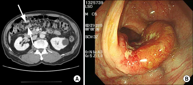
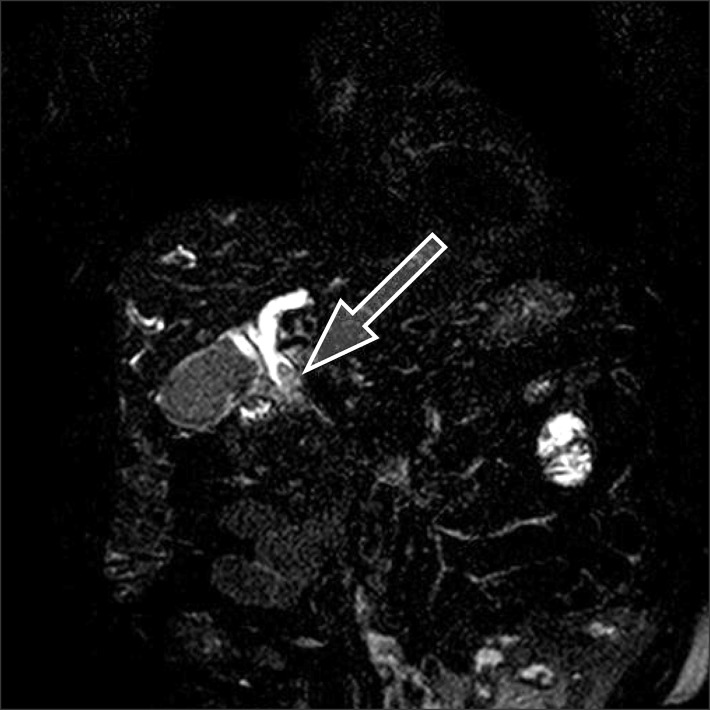
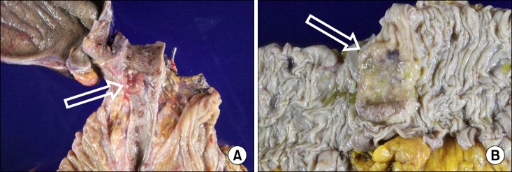
The patient was a 70-year-old male whose chief complaints were obstructive jaundice and weight loss. Abdominal imaging studies showed a 2.5 cm sized mass at the distal common bile duct, which was suggestive of bile duct cancer. Eccentric enhancing wall thickening in the transverse colon was also shown, suggesting concomitant colon cancer. A colonoscopy revealed a lumen-encircling ulcerofungating mass in the transverse colon, that was pathologically proven to be adenocarcinoma. The bile duct pathology was also adenocarcinoma. Pylorus-preserving pancreaticoduodenectomy and extended right hemicolectomy were performed under the diagnosis of double primary cancers. Postoperative histopathologic examination revealed moderately differentiated mucinous adenocarcinoma of transverse colon cancer, and mucinous adenocarcinoma of the distal common bile duct. Immunohistochemical staining studies showed that the bile duct cancer had metastasized from the colon cancer. The patient recovered uneventfully from surgery and will be undergoing chemotherapy for three months.

 求助内容:
求助内容: 应助结果提醒方式:


