Audrius Šileikis, Vitalija Nutautienė, Dmitrij Šeinin, Kęstutis Strupas
{"title":"胰腺实性假乳头状瘤7例分析。","authors":"Audrius Šileikis, Vitalija Nutautienė, Dmitrij Šeinin, Kęstutis Strupas","doi":"10.1159/000362183","DOIUrl":null,"url":null,"abstract":"<p><strong>Background: </strong>The purpose of this study was to describe as well as compare our surgical treatment experiences of solid pseudopapillary neoplasms (SPN) of the pancreas and to provide a review of the literature.</p><p><strong>Methods: </strong>A retrospective analysis of data from Vilnius University Hospital Santariskiu Klinikos (VUH SK) and of the literature, which was researched using Karger Publishers, Springer Science, BioMed Central, and disserCat databases, was conducted.</p><p><strong>Results: </strong>From 2001 to 2012, seven cases were identified with pathologically confirmed SPN diagnosis. A precise preoperative diagnosis was made by computertomography and magnetic resonance imaging. The median diameter of the tumors was 6.36 cm (range 1.5-12 cm). Surgical treatment was undertaken for all patients. Results of the immunohistochemical analysis confirmed a nuclear accumulation of β-catenin. The Ki-67 level was 1-2% in all of the cases. According to our collected data, all types of histological analysis revealed decent prognostic behavior with low mitotic activity (1-2 mitoses per 50 high power fields). Besides, angioinvasion, perineural invasion, and outside capsule invasion were not detected.</p><p><strong>Conclusions: </strong>There was no correlation between more aggressive types of SPN and tumor size, localization, age, and gender.</p>","PeriodicalId":49114,"journal":{"name":"Viszeralmedizin","volume":"30 3","pages":"211-5"},"PeriodicalIF":0.0000,"publicationDate":"2014-06-01","publicationTypes":"Journal Article","fieldsOfStudy":null,"isOpenAccess":false,"openAccessPdf":"https://sci-hub-pdf.com/10.1159/000362183","citationCount":"2","resultStr":"{\"title\":\"Solid Pseudopapillary Neoplasm of the Pancreas: Analysis of Seven Cases.\",\"authors\":\"Audrius Šileikis, Vitalija Nutautienė, Dmitrij Šeinin, Kęstutis Strupas\",\"doi\":\"10.1159/000362183\",\"DOIUrl\":null,\"url\":null,\"abstract\":\"<p><strong>Background: </strong>The purpose of this study was to describe as well as compare our surgical treatment experiences of solid pseudopapillary neoplasms (SPN) of the pancreas and to provide a review of the literature.</p><p><strong>Methods: </strong>A retrospective analysis of data from Vilnius University Hospital Santariskiu Klinikos (VUH SK) and of the literature, which was researched using Karger Publishers, Springer Science, BioMed Central, and disserCat databases, was conducted.</p><p><strong>Results: </strong>From 2001 to 2012, seven cases were identified with pathologically confirmed SPN diagnosis. A precise preoperative diagnosis was made by computertomography and magnetic resonance imaging. The median diameter of the tumors was 6.36 cm (range 1.5-12 cm). Surgical treatment was undertaken for all patients. Results of the immunohistochemical analysis confirmed a nuclear accumulation of β-catenin. The Ki-67 level was 1-2% in all of the cases. According to our collected data, all types of histological analysis revealed decent prognostic behavior with low mitotic activity (1-2 mitoses per 50 high power fields). Besides, angioinvasion, perineural invasion, and outside capsule invasion were not detected.</p><p><strong>Conclusions: </strong>There was no correlation between more aggressive types of SPN and tumor size, localization, age, and gender.</p>\",\"PeriodicalId\":49114,\"journal\":{\"name\":\"Viszeralmedizin\",\"volume\":\"30 3\",\"pages\":\"211-5\"},\"PeriodicalIF\":0.0000,\"publicationDate\":\"2014-06-01\",\"publicationTypes\":\"Journal Article\",\"fieldsOfStudy\":null,\"isOpenAccess\":false,\"openAccessPdf\":\"https://sci-hub-pdf.com/10.1159/000362183\",\"citationCount\":\"2\",\"resultStr\":null,\"platform\":\"Semanticscholar\",\"paperid\":null,\"PeriodicalName\":\"Viszeralmedizin\",\"FirstCategoryId\":\"1085\",\"ListUrlMain\":\"https://doi.org/10.1159/000362183\",\"RegionNum\":0,\"RegionCategory\":null,\"ArticlePicture\":[],\"TitleCN\":null,\"AbstractTextCN\":null,\"PMCID\":null,\"EPubDate\":\"\",\"PubModel\":\"\",\"JCR\":\"\",\"JCRName\":\"\",\"Score\":null,\"Total\":0}","platform":"Semanticscholar","paperid":null,"PeriodicalName":"Viszeralmedizin","FirstCategoryId":"1085","ListUrlMain":"https://doi.org/10.1159/000362183","RegionNum":0,"RegionCategory":null,"ArticlePicture":[],"TitleCN":null,"AbstractTextCN":null,"PMCID":null,"EPubDate":"","PubModel":"","JCR":"","JCRName":"","Score":null,"Total":0}
Solid Pseudopapillary Neoplasm of the Pancreas: Analysis of Seven Cases.
Background: The purpose of this study was to describe as well as compare our surgical treatment experiences of solid pseudopapillary neoplasms (SPN) of the pancreas and to provide a review of the literature.
Methods: A retrospective analysis of data from Vilnius University Hospital Santariskiu Klinikos (VUH SK) and of the literature, which was researched using Karger Publishers, Springer Science, BioMed Central, and disserCat databases, was conducted.
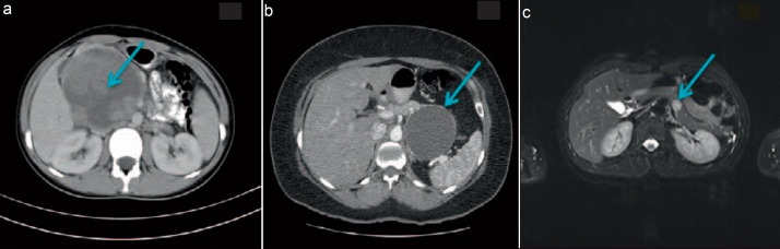
Results: From 2001 to 2012, seven cases were identified with pathologically confirmed SPN diagnosis. A precise preoperative diagnosis was made by computertomography and magnetic resonance imaging. The median diameter of the tumors was 6.36 cm (range 1.5-12 cm). Surgical treatment was undertaken for all patients. Results of the immunohistochemical analysis confirmed a nuclear accumulation of β-catenin. The Ki-67 level was 1-2% in all of the cases. According to our collected data, all types of histological analysis revealed decent prognostic behavior with low mitotic activity (1-2 mitoses per 50 high power fields). Besides, angioinvasion, perineural invasion, and outside capsule invasion were not detected.
Conclusions: There was no correlation between more aggressive types of SPN and tumor size, localization, age, and gender.
 求助内容:
求助内容: 应助结果提醒方式:


