Renata Siciliani Scalco, Stefen Brady, Jefferson Becker, Irenio Gomes, Janice L Holton, Henrique L Staub
{"title":"致编辑的信非典型肉芽肿性肌炎和肺结节病。","authors":"Renata Siciliani Scalco, Stefen Brady, Jefferson Becker, Irenio Gomes, Janice L Holton, Henrique L Staub","doi":"10.2174/1874312901409010057","DOIUrl":null,"url":null,"abstract":"A 45-year-old Brazilian white female was admitted with a four-year history of progressive proximal muscle weakness and myalgia. A chest radiogram was normal at the beginning of the clinical presentation. A few weeks before hospital admission, she developed diffuse weakness, dysphagia and exertion dyspnea. A physical examination showed weakness of facial and proximal muscles and severe wasting of hand muscles. Her family history was negative for neuromuscular diseases. The erythrocyte sedimentation rate was 92 mm in the first hour, and the serum creatine kinase level was high (6,683 IU/L, with a normal range being up to 176 IU/L). Antinuclear antibodies (1/640, granular pattern) and circulating anti-Ro/SSA antibodies (56.7 IU) were present. No other autoantibodies were found. A dried blood spot test for Pompe disease was negative. Neurophysiology assessment showed myopathic changes. A muscle biopsy performed 4 years after disease onset showed increased variation in fibre size, increased connective tissue, internal nuclei, occasional atrophic fibres and necrotic fibres. There was a prominent endomysial and perimysial inflammatory infiltrate composed of lymphoctes and macrophages with the formation of non-necrotic granulomas including small numbers of multinucleate giant cells (Fig. 1). A second piece of tissue taken from the same muscle showed no inflammatory features. Investigations for tuberculosis, fungi and brucellosis were negative. Lung computed tomography performed after 4 years of disease onset showed typical bilateral hilar lymphadenopathy and interstitial pneumonitis","PeriodicalId":39124,"journal":{"name":"Open Rheumatology Journal","volume":"9 ","pages":"57-9"},"PeriodicalIF":0.0000,"publicationDate":"2015-07-10","publicationTypes":"Journal Article","fieldsOfStudy":null,"isOpenAccess":false,"openAccessPdf":"https://ftp.ncbi.nlm.nih.gov/pub/pmc/oa_pdf/f3/c2/TORJ-9-57.PMC4541420.pdf","citationCount":"4","resultStr":"{\"title\":\"LETTER TO THE EDITOR Atypical Granulomatous Myositis and Pulmonary Sarcoidosis.\",\"authors\":\"Renata Siciliani Scalco, Stefen Brady, Jefferson Becker, Irenio Gomes, Janice L Holton, Henrique L Staub\",\"doi\":\"10.2174/1874312901409010057\",\"DOIUrl\":null,\"url\":null,\"abstract\":\"A 45-year-old Brazilian white female was admitted with a four-year history of progressive proximal muscle weakness and myalgia. A chest radiogram was normal at the beginning of the clinical presentation. A few weeks before hospital admission, she developed diffuse weakness, dysphagia and exertion dyspnea. A physical examination showed weakness of facial and proximal muscles and severe wasting of hand muscles. Her family history was negative for neuromuscular diseases. The erythrocyte sedimentation rate was 92 mm in the first hour, and the serum creatine kinase level was high (6,683 IU/L, with a normal range being up to 176 IU/L). Antinuclear antibodies (1/640, granular pattern) and circulating anti-Ro/SSA antibodies (56.7 IU) were present. No other autoantibodies were found. A dried blood spot test for Pompe disease was negative. Neurophysiology assessment showed myopathic changes. A muscle biopsy performed 4 years after disease onset showed increased variation in fibre size, increased connective tissue, internal nuclei, occasional atrophic fibres and necrotic fibres. There was a prominent endomysial and perimysial inflammatory infiltrate composed of lymphoctes and macrophages with the formation of non-necrotic granulomas including small numbers of multinucleate giant cells (Fig. 1). A second piece of tissue taken from the same muscle showed no inflammatory features. Investigations for tuberculosis, fungi and brucellosis were negative. Lung computed tomography performed after 4 years of disease onset showed typical bilateral hilar lymphadenopathy and interstitial pneumonitis\",\"PeriodicalId\":39124,\"journal\":{\"name\":\"Open Rheumatology Journal\",\"volume\":\"9 \",\"pages\":\"57-9\"},\"PeriodicalIF\":0.0000,\"publicationDate\":\"2015-07-10\",\"publicationTypes\":\"Journal Article\",\"fieldsOfStudy\":null,\"isOpenAccess\":false,\"openAccessPdf\":\"https://ftp.ncbi.nlm.nih.gov/pub/pmc/oa_pdf/f3/c2/TORJ-9-57.PMC4541420.pdf\",\"citationCount\":\"4\",\"resultStr\":null,\"platform\":\"Semanticscholar\",\"paperid\":null,\"PeriodicalName\":\"Open Rheumatology Journal\",\"FirstCategoryId\":\"1085\",\"ListUrlMain\":\"https://doi.org/10.2174/1874312901409010057\",\"RegionNum\":0,\"RegionCategory\":null,\"ArticlePicture\":[],\"TitleCN\":null,\"AbstractTextCN\":null,\"PMCID\":null,\"EPubDate\":\"2015/1/1 0:00:00\",\"PubModel\":\"eCollection\",\"JCR\":\"Q4\",\"JCRName\":\"Medicine\",\"Score\":null,\"Total\":0}","platform":"Semanticscholar","paperid":null,"PeriodicalName":"Open Rheumatology Journal","FirstCategoryId":"1085","ListUrlMain":"https://doi.org/10.2174/1874312901409010057","RegionNum":0,"RegionCategory":null,"ArticlePicture":[],"TitleCN":null,"AbstractTextCN":null,"PMCID":null,"EPubDate":"2015/1/1 0:00:00","PubModel":"eCollection","JCR":"Q4","JCRName":"Medicine","Score":null,"Total":0}
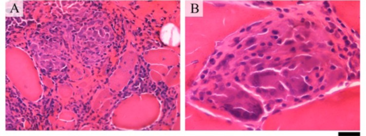
LETTER TO THE EDITOR Atypical Granulomatous Myositis and Pulmonary Sarcoidosis.
A 45-year-old Brazilian white female was admitted with a four-year history of progressive proximal muscle weakness and myalgia. A chest radiogram was normal at the beginning of the clinical presentation. A few weeks before hospital admission, she developed diffuse weakness, dysphagia and exertion dyspnea. A physical examination showed weakness of facial and proximal muscles and severe wasting of hand muscles. Her family history was negative for neuromuscular diseases. The erythrocyte sedimentation rate was 92 mm in the first hour, and the serum creatine kinase level was high (6,683 IU/L, with a normal range being up to 176 IU/L). Antinuclear antibodies (1/640, granular pattern) and circulating anti-Ro/SSA antibodies (56.7 IU) were present. No other autoantibodies were found. A dried blood spot test for Pompe disease was negative. Neurophysiology assessment showed myopathic changes. A muscle biopsy performed 4 years after disease onset showed increased variation in fibre size, increased connective tissue, internal nuclei, occasional atrophic fibres and necrotic fibres. There was a prominent endomysial and perimysial inflammatory infiltrate composed of lymphoctes and macrophages with the formation of non-necrotic granulomas including small numbers of multinucleate giant cells (Fig. 1). A second piece of tissue taken from the same muscle showed no inflammatory features. Investigations for tuberculosis, fungi and brucellosis were negative. Lung computed tomography performed after 4 years of disease onset showed typical bilateral hilar lymphadenopathy and interstitial pneumonitis
期刊介绍:
ENTHAM Open publishes a number of peer-reviewed, open access journals. These free-to-view online journals cover all major disciplines of science, medicine, technology and social sciences. BENTHAM Open provides researchers a platform to rapidly publish their research in a good-quality peer-reviewed journal. All peer-reviewed accepted submissions meeting high research and ethical standards are published with free access to all.

 求助内容:
求助内容: 应助结果提醒方式:


