{"title":"偶发性胰腺囊性病变的处理。","authors":"Christian Jenssen, Stefan Kahl","doi":"10.1159/000375282","DOIUrl":null,"url":null,"abstract":"<p><strong>Background: </strong>Pancreatic cystic lesions (PCL) are common. They are increasingly detected as an incidental finding of transabdominal ultrasound or cross-sectional imaging. In contrast to other parenchymal organs, dysontogenetic pancreatic cysts are extremely rare. In symptomatic patients the most frequent PCL are acute and chronic pseudocysts. The majority of incidental cystic lesions, however, are neoplasias which have different risks of malignancy.</p><p><strong>Methods: </strong>PubMed was searched for studies, reviews, meta-analyses, and guidelines using the following key words: ('pancreatic cystic lesions' OR 'cystic pancreatic lesions' OR 'intraductal papillary mucinous neoplasia' OR 'mucinous cystic neoplasia' OR 'pancreatic cyst' OR 'pancreatic pseudocyst') AND (management OR treatment OR outcome OR prognosis OR diagnosis OR imaging OR 'endoscopic ultrasound' EUS-FNA OR EUS OR 'endoscopic ultrasonography' OR CT OR MRI). Retrieved papers were reviewed with regard to the diagnostic and therapeutic management of incidental PCL.</p><p><strong>Results: </strong>In addition to clinical criteria, transabdominal ultrasonography including contrast-enhanced ultrasonography, cross-sectional radiological imaging, and endoscopic ultrasound (EUS) are used for diagnostic characterization and risk assessment. EUS plays an outstanding role in differential diagnosis and prognostic characterization of incidental PCL. In a single examination it is possible to perform high-resolution morphological description, perfusion imaging, as well as fine-needle aspiration of cyst content, cyst wall, and solid components. An international consensus guideline has defined worrisome and high-risk criteria for the risk assessment of mucinous pancreatic cysts, which are mainly based on the results of EUS and cross-sectional imaging. Nevertheless, despite diagnostic progress and guideline recommendations, differential diagnosis and management decisions remain difficult. This review will discuss problems in and approaches to the diagnosis of incidental PCL.</p><p><strong>Conclusion: </strong>An evidence-based algorithm for the diagnosis of incidental PCL is proposed.</p>","PeriodicalId":49114,"journal":{"name":"Viszeralmedizin","volume":"31 1","pages":"14-24"},"PeriodicalIF":0.0000,"publicationDate":"2015-02-01","publicationTypes":"Journal Article","fieldsOfStudy":null,"isOpenAccess":false,"openAccessPdf":"https://sci-hub-pdf.com/10.1159/000375282","citationCount":"20","resultStr":"{\"title\":\"Management of Incidental Pancreatic Cystic Lesions.\",\"authors\":\"Christian Jenssen, Stefan Kahl\",\"doi\":\"10.1159/000375282\",\"DOIUrl\":null,\"url\":null,\"abstract\":\"<p><strong>Background: </strong>Pancreatic cystic lesions (PCL) are common. They are increasingly detected as an incidental finding of transabdominal ultrasound or cross-sectional imaging. In contrast to other parenchymal organs, dysontogenetic pancreatic cysts are extremely rare. In symptomatic patients the most frequent PCL are acute and chronic pseudocysts. The majority of incidental cystic lesions, however, are neoplasias which have different risks of malignancy.</p><p><strong>Methods: </strong>PubMed was searched for studies, reviews, meta-analyses, and guidelines using the following key words: ('pancreatic cystic lesions' OR 'cystic pancreatic lesions' OR 'intraductal papillary mucinous neoplasia' OR 'mucinous cystic neoplasia' OR 'pancreatic cyst' OR 'pancreatic pseudocyst') AND (management OR treatment OR outcome OR prognosis OR diagnosis OR imaging OR 'endoscopic ultrasound' EUS-FNA OR EUS OR 'endoscopic ultrasonography' OR CT OR MRI). Retrieved papers were reviewed with regard to the diagnostic and therapeutic management of incidental PCL.</p><p><strong>Results: </strong>In addition to clinical criteria, transabdominal ultrasonography including contrast-enhanced ultrasonography, cross-sectional radiological imaging, and endoscopic ultrasound (EUS) are used for diagnostic characterization and risk assessment. EUS plays an outstanding role in differential diagnosis and prognostic characterization of incidental PCL. In a single examination it is possible to perform high-resolution morphological description, perfusion imaging, as well as fine-needle aspiration of cyst content, cyst wall, and solid components. An international consensus guideline has defined worrisome and high-risk criteria for the risk assessment of mucinous pancreatic cysts, which are mainly based on the results of EUS and cross-sectional imaging. Nevertheless, despite diagnostic progress and guideline recommendations, differential diagnosis and management decisions remain difficult. This review will discuss problems in and approaches to the diagnosis of incidental PCL.</p><p><strong>Conclusion: </strong>An evidence-based algorithm for the diagnosis of incidental PCL is proposed.</p>\",\"PeriodicalId\":49114,\"journal\":{\"name\":\"Viszeralmedizin\",\"volume\":\"31 1\",\"pages\":\"14-24\"},\"PeriodicalIF\":0.0000,\"publicationDate\":\"2015-02-01\",\"publicationTypes\":\"Journal Article\",\"fieldsOfStudy\":null,\"isOpenAccess\":false,\"openAccessPdf\":\"https://sci-hub-pdf.com/10.1159/000375282\",\"citationCount\":\"20\",\"resultStr\":null,\"platform\":\"Semanticscholar\",\"paperid\":null,\"PeriodicalName\":\"Viszeralmedizin\",\"FirstCategoryId\":\"1085\",\"ListUrlMain\":\"https://doi.org/10.1159/000375282\",\"RegionNum\":0,\"RegionCategory\":null,\"ArticlePicture\":[],\"TitleCN\":null,\"AbstractTextCN\":null,\"PMCID\":null,\"EPubDate\":\"\",\"PubModel\":\"\",\"JCR\":\"\",\"JCRName\":\"\",\"Score\":null,\"Total\":0}","platform":"Semanticscholar","paperid":null,"PeriodicalName":"Viszeralmedizin","FirstCategoryId":"1085","ListUrlMain":"https://doi.org/10.1159/000375282","RegionNum":0,"RegionCategory":null,"ArticlePicture":[],"TitleCN":null,"AbstractTextCN":null,"PMCID":null,"EPubDate":"","PubModel":"","JCR":"","JCRName":"","Score":null,"Total":0}
Management of Incidental Pancreatic Cystic Lesions.
Background: Pancreatic cystic lesions (PCL) are common. They are increasingly detected as an incidental finding of transabdominal ultrasound or cross-sectional imaging. In contrast to other parenchymal organs, dysontogenetic pancreatic cysts are extremely rare. In symptomatic patients the most frequent PCL are acute and chronic pseudocysts. The majority of incidental cystic lesions, however, are neoplasias which have different risks of malignancy.
Methods: PubMed was searched for studies, reviews, meta-analyses, and guidelines using the following key words: ('pancreatic cystic lesions' OR 'cystic pancreatic lesions' OR 'intraductal papillary mucinous neoplasia' OR 'mucinous cystic neoplasia' OR 'pancreatic cyst' OR 'pancreatic pseudocyst') AND (management OR treatment OR outcome OR prognosis OR diagnosis OR imaging OR 'endoscopic ultrasound' EUS-FNA OR EUS OR 'endoscopic ultrasonography' OR CT OR MRI). Retrieved papers were reviewed with regard to the diagnostic and therapeutic management of incidental PCL.
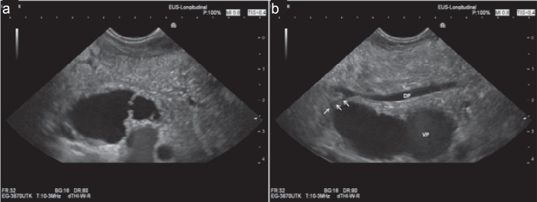
Results: In addition to clinical criteria, transabdominal ultrasonography including contrast-enhanced ultrasonography, cross-sectional radiological imaging, and endoscopic ultrasound (EUS) are used for diagnostic characterization and risk assessment. EUS plays an outstanding role in differential diagnosis and prognostic characterization of incidental PCL. In a single examination it is possible to perform high-resolution morphological description, perfusion imaging, as well as fine-needle aspiration of cyst content, cyst wall, and solid components. An international consensus guideline has defined worrisome and high-risk criteria for the risk assessment of mucinous pancreatic cysts, which are mainly based on the results of EUS and cross-sectional imaging. Nevertheless, despite diagnostic progress and guideline recommendations, differential diagnosis and management decisions remain difficult. This review will discuss problems in and approaches to the diagnosis of incidental PCL.
Conclusion: An evidence-based algorithm for the diagnosis of incidental PCL is proposed.
 求助内容:
求助内容: 应助结果提醒方式:


