Cheon-Soo Park, Shin Hwang, Dong-Hwan Jung, Gi-Won Song, Deok-Bog Moon, Chul-Soo Ahn, Gil-Chun Park, Ki-Hun Kim, Tae-Yong Ha, Sung-Gyu Lee
{"title":"成人活体肝移植后肠性肺肿3例报告并文献复习。","authors":"Cheon-Soo Park, Shin Hwang, Dong-Hwan Jung, Gi-Won Song, Deok-Bog Moon, Chul-Soo Ahn, Gil-Chun Park, Ki-Hun Kim, Tae-Yong Ha, Sung-Gyu Lee","doi":"10.14701/kjhbps.2015.19.1.25","DOIUrl":null,"url":null,"abstract":"<p><strong>Backgrounds/aims: </strong>Pneumatosis intestinalis (PI) is a condition in which multiple gas-filled mural cysts develop in the gastrointestinal tract. Although its exact etiology remains obscure, PI is rarely observed in liver transplant (LT) recipients.</p><p><strong>Methods: </strong>In 317 cases of adult living donor LT (LDLT) performed during 2011, PI developed in three patients during the 3 year follow-up.</p><p><strong>Results: </strong>Of these three patients, the two who demonstrated PI at 6 weeks and 2 months after LT, respectively, were asymptomatic and showed no signs of secondary complications. Diagnosis was made incidentally using abdominal radiographs and computed tomography (CT) scans. PI was identified in the right ascending colon with concomitant pneumoperitoneum. These two patients received supportive care and maintained a regular diet. Follow-up CT scans demonstrated spontaneous resolution of PI with no complications. The third patient was admitted to the emergency room 30 months after LDLT. His symptoms included poor oral intake and intermittent abdominal pain with no passage of gas. Abdominal radiography and CT scans demonstrated PI in the entire small bowel, with small bowel dilatation, pneumoperitoneum, and pneumoretroperitoneum, but no peritonitis. Physical examination revealed abdominal distension but no tenderness or rebound tenderness. After 1 week of conservative treatment, including bowel rest and antibiotics therapy, PI and pneumoperitoneum resolved spontaneously without complications.</p><p><strong>Conclusions: </strong>We suggest that adult LDLT recipients who develop asymptomatic or symptomatic PI with no signs of secondary complications can be successfully managed with conservative treatment.</p>","PeriodicalId":91136,"journal":{"name":"Korean journal of hepato-biliary-pancreatic surgery","volume":"19 1","pages":"25-9"},"PeriodicalIF":0.0000,"publicationDate":"2015-02-01","publicationTypes":"Journal Article","fieldsOfStudy":null,"isOpenAccess":false,"openAccessPdf":"https://sci-hub-pdf.com/10.14701/kjhbps.2015.19.1.25","citationCount":"8","resultStr":"{\"title\":\"Pneumatosis intestinalis after adult living donor liver transplantation: report of three cases and collective literature review.\",\"authors\":\"Cheon-Soo Park, Shin Hwang, Dong-Hwan Jung, Gi-Won Song, Deok-Bog Moon, Chul-Soo Ahn, Gil-Chun Park, Ki-Hun Kim, Tae-Yong Ha, Sung-Gyu Lee\",\"doi\":\"10.14701/kjhbps.2015.19.1.25\",\"DOIUrl\":null,\"url\":null,\"abstract\":\"<p><strong>Backgrounds/aims: </strong>Pneumatosis intestinalis (PI) is a condition in which multiple gas-filled mural cysts develop in the gastrointestinal tract. Although its exact etiology remains obscure, PI is rarely observed in liver transplant (LT) recipients.</p><p><strong>Methods: </strong>In 317 cases of adult living donor LT (LDLT) performed during 2011, PI developed in three patients during the 3 year follow-up.</p><p><strong>Results: </strong>Of these three patients, the two who demonstrated PI at 6 weeks and 2 months after LT, respectively, were asymptomatic and showed no signs of secondary complications. Diagnosis was made incidentally using abdominal radiographs and computed tomography (CT) scans. PI was identified in the right ascending colon with concomitant pneumoperitoneum. These two patients received supportive care and maintained a regular diet. Follow-up CT scans demonstrated spontaneous resolution of PI with no complications. The third patient was admitted to the emergency room 30 months after LDLT. His symptoms included poor oral intake and intermittent abdominal pain with no passage of gas. Abdominal radiography and CT scans demonstrated PI in the entire small bowel, with small bowel dilatation, pneumoperitoneum, and pneumoretroperitoneum, but no peritonitis. Physical examination revealed abdominal distension but no tenderness or rebound tenderness. After 1 week of conservative treatment, including bowel rest and antibiotics therapy, PI and pneumoperitoneum resolved spontaneously without complications.</p><p><strong>Conclusions: </strong>We suggest that adult LDLT recipients who develop asymptomatic or symptomatic PI with no signs of secondary complications can be successfully managed with conservative treatment.</p>\",\"PeriodicalId\":91136,\"journal\":{\"name\":\"Korean journal of hepato-biliary-pancreatic surgery\",\"volume\":\"19 1\",\"pages\":\"25-9\"},\"PeriodicalIF\":0.0000,\"publicationDate\":\"2015-02-01\",\"publicationTypes\":\"Journal Article\",\"fieldsOfStudy\":null,\"isOpenAccess\":false,\"openAccessPdf\":\"https://sci-hub-pdf.com/10.14701/kjhbps.2015.19.1.25\",\"citationCount\":\"8\",\"resultStr\":null,\"platform\":\"Semanticscholar\",\"paperid\":null,\"PeriodicalName\":\"Korean journal of hepato-biliary-pancreatic surgery\",\"FirstCategoryId\":\"1085\",\"ListUrlMain\":\"https://doi.org/10.14701/kjhbps.2015.19.1.25\",\"RegionNum\":0,\"RegionCategory\":null,\"ArticlePicture\":[],\"TitleCN\":null,\"AbstractTextCN\":null,\"PMCID\":null,\"EPubDate\":\"2015/2/28 0:00:00\",\"PubModel\":\"Epub\",\"JCR\":\"\",\"JCRName\":\"\",\"Score\":null,\"Total\":0}","platform":"Semanticscholar","paperid":null,"PeriodicalName":"Korean journal of hepato-biliary-pancreatic surgery","FirstCategoryId":"1085","ListUrlMain":"https://doi.org/10.14701/kjhbps.2015.19.1.25","RegionNum":0,"RegionCategory":null,"ArticlePicture":[],"TitleCN":null,"AbstractTextCN":null,"PMCID":null,"EPubDate":"2015/2/28 0:00:00","PubModel":"Epub","JCR":"","JCRName":"","Score":null,"Total":0}
Pneumatosis intestinalis after adult living donor liver transplantation: report of three cases and collective literature review.
Backgrounds/aims: Pneumatosis intestinalis (PI) is a condition in which multiple gas-filled mural cysts develop in the gastrointestinal tract. Although its exact etiology remains obscure, PI is rarely observed in liver transplant (LT) recipients.
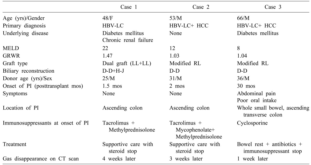
Methods: In 317 cases of adult living donor LT (LDLT) performed during 2011, PI developed in three patients during the 3 year follow-up.
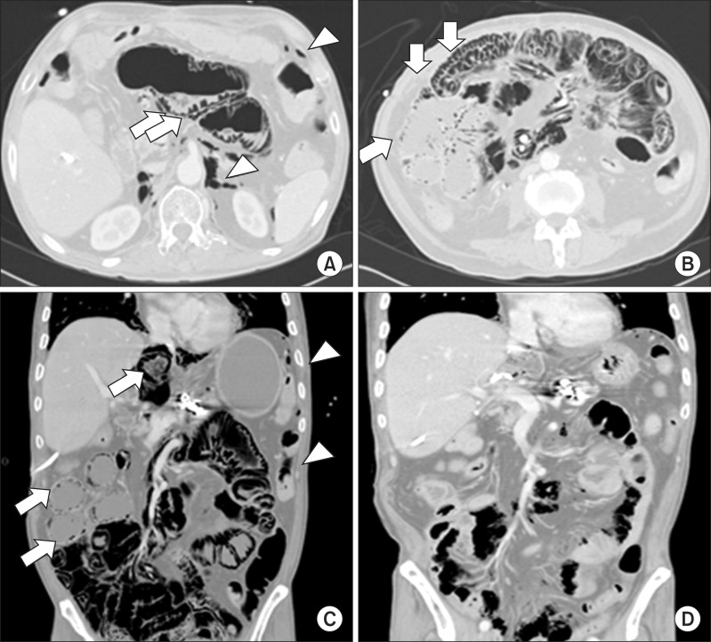
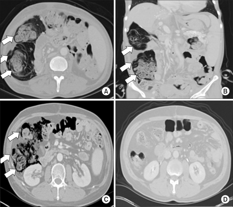
Results: Of these three patients, the two who demonstrated PI at 6 weeks and 2 months after LT, respectively, were asymptomatic and showed no signs of secondary complications. Diagnosis was made incidentally using abdominal radiographs and computed tomography (CT) scans. PI was identified in the right ascending colon with concomitant pneumoperitoneum. These two patients received supportive care and maintained a regular diet. Follow-up CT scans demonstrated spontaneous resolution of PI with no complications. The third patient was admitted to the emergency room 30 months after LDLT. His symptoms included poor oral intake and intermittent abdominal pain with no passage of gas. Abdominal radiography and CT scans demonstrated PI in the entire small bowel, with small bowel dilatation, pneumoperitoneum, and pneumoretroperitoneum, but no peritonitis. Physical examination revealed abdominal distension but no tenderness or rebound tenderness. After 1 week of conservative treatment, including bowel rest and antibiotics therapy, PI and pneumoperitoneum resolved spontaneously without complications.
Conclusions: We suggest that adult LDLT recipients who develop asymptomatic or symptomatic PI with no signs of secondary complications can be successfully managed with conservative treatment.

 求助内容:
求助内容: 应助结果提醒方式:


