Younguk Kim, Guen Young Lee, Sujin Kim, Kwang-Sup Song, Hee Sung Kim
{"title":"马尾原发性t细胞淋巴瘤的MR影像学特征:1例报告及文献复习。","authors":"Younguk Kim, Guen Young Lee, Sujin Kim, Kwang-Sup Song, Hee Sung Kim","doi":"10.3348/jksr.2020.0195","DOIUrl":null,"url":null,"abstract":"<p><p>Primary central nervous system lymphoma is a rare form of extranodal non-Hodgkin lymphoma, and primary T-cell lymphoma of the cauda equina is extremely rare. We describe a case involving a 56-year-old female who presented with low back pain and radiating leg pain for 4 months. MRI of the lumbar spine revealed an elongated, multinodular intradural lesion of approximately 10 cm from the L4 body to the S2 body level with iso-signal intensity on T1-weighted imaging, heterogeneous iso- and high-signal intensity on T2-weighted imaging, and a heterogeneous intense enhancement on gadolinium contrast-enhanced T1-weighted imaging. A peripheral T-cell lymphoma of the cauda equina was diagnosed on the basis of immunohistochemical and T-cell receptor gamma gene rearrangement analysis after intradural biopsy of the mass.</p>","PeriodicalId":74904,"journal":{"name":"Taehan Yongsang Uihakhoe chi","volume":"82 6","pages":"1613-1618"},"PeriodicalIF":0.0000,"publicationDate":"2021-11-01","publicationTypes":"Journal Article","fieldsOfStudy":null,"isOpenAccess":false,"openAccessPdf":"https://ftp.ncbi.nlm.nih.gov/pub/pmc/oa_pdf/60/a1/jksr-82-1613.PMC9431974.pdf","citationCount":"0","resultStr":"{\"title\":\"MR Imaging Characteristics of Primary T-Cell Lymphoma of the Cauda Equina: A Case Report and Literature Review.\",\"authors\":\"Younguk Kim, Guen Young Lee, Sujin Kim, Kwang-Sup Song, Hee Sung Kim\",\"doi\":\"10.3348/jksr.2020.0195\",\"DOIUrl\":null,\"url\":null,\"abstract\":\"<p><p>Primary central nervous system lymphoma is a rare form of extranodal non-Hodgkin lymphoma, and primary T-cell lymphoma of the cauda equina is extremely rare. We describe a case involving a 56-year-old female who presented with low back pain and radiating leg pain for 4 months. MRI of the lumbar spine revealed an elongated, multinodular intradural lesion of approximately 10 cm from the L4 body to the S2 body level with iso-signal intensity on T1-weighted imaging, heterogeneous iso- and high-signal intensity on T2-weighted imaging, and a heterogeneous intense enhancement on gadolinium contrast-enhanced T1-weighted imaging. A peripheral T-cell lymphoma of the cauda equina was diagnosed on the basis of immunohistochemical and T-cell receptor gamma gene rearrangement analysis after intradural biopsy of the mass.</p>\",\"PeriodicalId\":74904,\"journal\":{\"name\":\"Taehan Yongsang Uihakhoe chi\",\"volume\":\"82 6\",\"pages\":\"1613-1618\"},\"PeriodicalIF\":0.0000,\"publicationDate\":\"2021-11-01\",\"publicationTypes\":\"Journal Article\",\"fieldsOfStudy\":null,\"isOpenAccess\":false,\"openAccessPdf\":\"https://ftp.ncbi.nlm.nih.gov/pub/pmc/oa_pdf/60/a1/jksr-82-1613.PMC9431974.pdf\",\"citationCount\":\"0\",\"resultStr\":null,\"platform\":\"Semanticscholar\",\"paperid\":null,\"PeriodicalName\":\"Taehan Yongsang Uihakhoe chi\",\"FirstCategoryId\":\"1085\",\"ListUrlMain\":\"https://doi.org/10.3348/jksr.2020.0195\",\"RegionNum\":0,\"RegionCategory\":null,\"ArticlePicture\":[],\"TitleCN\":null,\"AbstractTextCN\":null,\"PMCID\":null,\"EPubDate\":\"2021/10/18 0:00:00\",\"PubModel\":\"Epub\",\"JCR\":\"\",\"JCRName\":\"\",\"Score\":null,\"Total\":0}","platform":"Semanticscholar","paperid":null,"PeriodicalName":"Taehan Yongsang Uihakhoe chi","FirstCategoryId":"1085","ListUrlMain":"https://doi.org/10.3348/jksr.2020.0195","RegionNum":0,"RegionCategory":null,"ArticlePicture":[],"TitleCN":null,"AbstractTextCN":null,"PMCID":null,"EPubDate":"2021/10/18 0:00:00","PubModel":"Epub","JCR":"","JCRName":"","Score":null,"Total":0}
MR Imaging Characteristics of Primary T-Cell Lymphoma of the Cauda Equina: A Case Report and Literature Review.
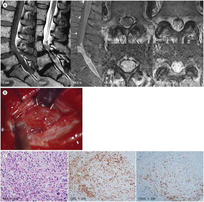
Primary central nervous system lymphoma is a rare form of extranodal non-Hodgkin lymphoma, and primary T-cell lymphoma of the cauda equina is extremely rare. We describe a case involving a 56-year-old female who presented with low back pain and radiating leg pain for 4 months. MRI of the lumbar spine revealed an elongated, multinodular intradural lesion of approximately 10 cm from the L4 body to the S2 body level with iso-signal intensity on T1-weighted imaging, heterogeneous iso- and high-signal intensity on T2-weighted imaging, and a heterogeneous intense enhancement on gadolinium contrast-enhanced T1-weighted imaging. A peripheral T-cell lymphoma of the cauda equina was diagnosed on the basis of immunohistochemical and T-cell receptor gamma gene rearrangement analysis after intradural biopsy of the mass.

 求助内容:
求助内容: 应助结果提醒方式:


