{"title":"婴儿中耳纤维性错构瘤的CT和MRI表现1例。","authors":"Sang Hun Baek, Sanghyeon Kim, Kyungjae Lim","doi":"10.3348/jksr.2021.0066","DOIUrl":null,"url":null,"abstract":"<p><p>Fibrous hamartoma of infancy in the middle ear is extremely rare. We report the case of a 26-month-old male patient who presented with a mass in the left middle ear. A temporal bone CT scan showed complete opacification of the left middle ear and mastoid air cells without ossicular erosion. On MRI, the mass revealed heterogeneous signal intensities indicative of fat and fibrous components. A definitive diagnosis was made postoperatively based on the histological results. Although rare, fibrous hamartoma of infancy should be considered as a differential diagnosis of a middle ear mass during childhood.</p>","PeriodicalId":74904,"journal":{"name":"Taehan Yongsang Uihakhoe chi","volume":" ","pages":"420-424"},"PeriodicalIF":0.0000,"publicationDate":"2022-03-01","publicationTypes":"Journal Article","fieldsOfStudy":null,"isOpenAccess":false,"openAccessPdf":"https://ftp.ncbi.nlm.nih.gov/pub/pmc/oa_pdf/78/b3/jksr-83-420.PMC9514445.pdf","citationCount":"0","resultStr":"{\"title\":\"CT and MRI Features of Middle Ear Fibrous Hamartoma of Infancy: A Case Report.\",\"authors\":\"Sang Hun Baek, Sanghyeon Kim, Kyungjae Lim\",\"doi\":\"10.3348/jksr.2021.0066\",\"DOIUrl\":null,\"url\":null,\"abstract\":\"<p><p>Fibrous hamartoma of infancy in the middle ear is extremely rare. We report the case of a 26-month-old male patient who presented with a mass in the left middle ear. A temporal bone CT scan showed complete opacification of the left middle ear and mastoid air cells without ossicular erosion. On MRI, the mass revealed heterogeneous signal intensities indicative of fat and fibrous components. A definitive diagnosis was made postoperatively based on the histological results. Although rare, fibrous hamartoma of infancy should be considered as a differential diagnosis of a middle ear mass during childhood.</p>\",\"PeriodicalId\":74904,\"journal\":{\"name\":\"Taehan Yongsang Uihakhoe chi\",\"volume\":\" \",\"pages\":\"420-424\"},\"PeriodicalIF\":0.0000,\"publicationDate\":\"2022-03-01\",\"publicationTypes\":\"Journal Article\",\"fieldsOfStudy\":null,\"isOpenAccess\":false,\"openAccessPdf\":\"https://ftp.ncbi.nlm.nih.gov/pub/pmc/oa_pdf/78/b3/jksr-83-420.PMC9514445.pdf\",\"citationCount\":\"0\",\"resultStr\":null,\"platform\":\"Semanticscholar\",\"paperid\":null,\"PeriodicalName\":\"Taehan Yongsang Uihakhoe chi\",\"FirstCategoryId\":\"1085\",\"ListUrlMain\":\"https://doi.org/10.3348/jksr.2021.0066\",\"RegionNum\":0,\"RegionCategory\":null,\"ArticlePicture\":[],\"TitleCN\":null,\"AbstractTextCN\":null,\"PMCID\":null,\"EPubDate\":\"2021/11/4 0:00:00\",\"PubModel\":\"Epub\",\"JCR\":\"\",\"JCRName\":\"\",\"Score\":null,\"Total\":0}","platform":"Semanticscholar","paperid":null,"PeriodicalName":"Taehan Yongsang Uihakhoe chi","FirstCategoryId":"1085","ListUrlMain":"https://doi.org/10.3348/jksr.2021.0066","RegionNum":0,"RegionCategory":null,"ArticlePicture":[],"TitleCN":null,"AbstractTextCN":null,"PMCID":null,"EPubDate":"2021/11/4 0:00:00","PubModel":"Epub","JCR":"","JCRName":"","Score":null,"Total":0}
CT and MRI Features of Middle Ear Fibrous Hamartoma of Infancy: A Case Report.
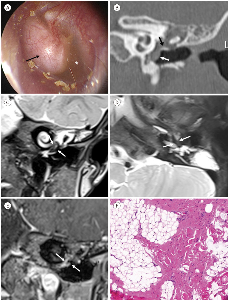
Fibrous hamartoma of infancy in the middle ear is extremely rare. We report the case of a 26-month-old male patient who presented with a mass in the left middle ear. A temporal bone CT scan showed complete opacification of the left middle ear and mastoid air cells without ossicular erosion. On MRI, the mass revealed heterogeneous signal intensities indicative of fat and fibrous components. A definitive diagnosis was made postoperatively based on the histological results. Although rare, fibrous hamartoma of infancy should be considered as a differential diagnosis of a middle ear mass during childhood.

 求助内容:
求助内容: 应助结果提醒方式:


