Hyung In Choi, Min Jeong Choi, Bong Man Kim, Hwan Namgung, Seung Kyu Choi
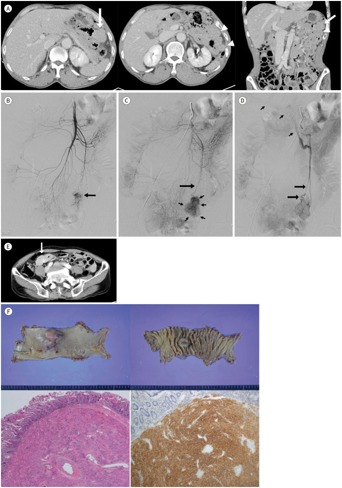
{"title":"鉴别小肠胃肠道间质瘤为隐蔽性胃肠道出血的罪魁祸首:强调血管造影结果。","authors":"Hyung In Choi, Min Jeong Choi, Bong Man Kim, Hwan Namgung, Seung Kyu Choi","doi":"10.3348/jksr.2021.0029","DOIUrl":null,"url":null,"abstract":"<p><p>Gastrointestinal stromal tumors (GISTs) are not uncommon and often cause gastrointestinal bleeding. GISTs occurring in the small intestine are occasionally difficult to identify by endoscopy and CT. In this case, the patient underwent CT three times before surgery, and the lesion was found to be located in a different area of the abdominal cavity on each CT scan. Moreover, the lesion was missed in the first two CT images because it was difficult to distinguish it from the nearby collapsed small intestine. The lesion was eventually detected through angiography; however, the correct diagnosis and treatment were delayed for 3 years because it was mistaken for a vascular malformation, which is the most common cause of obscure GI bleeding in elderly patients. This report emphasizes the need for interventional radiologists to be updated and vigilant of the angiographic features of GISTs to make an accurate diagnosis and establish a management strategy.</p>","PeriodicalId":74904,"journal":{"name":"Taehan Yongsang Uihakhoe chi","volume":" ","pages":"400-405"},"PeriodicalIF":0.0000,"publicationDate":"2022-03-01","publicationTypes":"Journal Article","fieldsOfStudy":null,"isOpenAccess":false,"openAccessPdf":"https://ftp.ncbi.nlm.nih.gov/pub/pmc/oa_pdf/8f/42/jksr-83-400.PMC9514442.pdf","citationCount":"0","resultStr":"{\"title\":\"Identifying Small Bowel Gastrointestinal Stromal Tumor as the Culprit Lesion in Obscure Gastrointestinal Bleeding: Emphasis on Angiographic Findings.\",\"authors\":\"Hyung In Choi, Min Jeong Choi, Bong Man Kim, Hwan Namgung, Seung Kyu Choi\",\"doi\":\"10.3348/jksr.2021.0029\",\"DOIUrl\":null,\"url\":null,\"abstract\":\"<p><p>Gastrointestinal stromal tumors (GISTs) are not uncommon and often cause gastrointestinal bleeding. GISTs occurring in the small intestine are occasionally difficult to identify by endoscopy and CT. In this case, the patient underwent CT three times before surgery, and the lesion was found to be located in a different area of the abdominal cavity on each CT scan. Moreover, the lesion was missed in the first two CT images because it was difficult to distinguish it from the nearby collapsed small intestine. The lesion was eventually detected through angiography; however, the correct diagnosis and treatment were delayed for 3 years because it was mistaken for a vascular malformation, which is the most common cause of obscure GI bleeding in elderly patients. This report emphasizes the need for interventional radiologists to be updated and vigilant of the angiographic features of GISTs to make an accurate diagnosis and establish a management strategy.</p>\",\"PeriodicalId\":74904,\"journal\":{\"name\":\"Taehan Yongsang Uihakhoe chi\",\"volume\":\" \",\"pages\":\"400-405\"},\"PeriodicalIF\":0.0000,\"publicationDate\":\"2022-03-01\",\"publicationTypes\":\"Journal Article\",\"fieldsOfStudy\":null,\"isOpenAccess\":false,\"openAccessPdf\":\"https://ftp.ncbi.nlm.nih.gov/pub/pmc/oa_pdf/8f/42/jksr-83-400.PMC9514442.pdf\",\"citationCount\":\"0\",\"resultStr\":null,\"platform\":\"Semanticscholar\",\"paperid\":null,\"PeriodicalName\":\"Taehan Yongsang Uihakhoe chi\",\"FirstCategoryId\":\"1085\",\"ListUrlMain\":\"https://doi.org/10.3348/jksr.2021.0029\",\"RegionNum\":0,\"RegionCategory\":null,\"ArticlePicture\":[],\"TitleCN\":null,\"AbstractTextCN\":null,\"PMCID\":null,\"EPubDate\":\"2021/11/4 0:00:00\",\"PubModel\":\"Epub\",\"JCR\":\"\",\"JCRName\":\"\",\"Score\":null,\"Total\":0}","platform":"Semanticscholar","paperid":null,"PeriodicalName":"Taehan Yongsang Uihakhoe chi","FirstCategoryId":"1085","ListUrlMain":"https://doi.org/10.3348/jksr.2021.0029","RegionNum":0,"RegionCategory":null,"ArticlePicture":[],"TitleCN":null,"AbstractTextCN":null,"PMCID":null,"EPubDate":"2021/11/4 0:00:00","PubModel":"Epub","JCR":"","JCRName":"","Score":null,"Total":0}
Identifying Small Bowel Gastrointestinal Stromal Tumor as the Culprit Lesion in Obscure Gastrointestinal Bleeding: Emphasis on Angiographic Findings.
Gastrointestinal stromal tumors (GISTs) are not uncommon and often cause gastrointestinal bleeding. GISTs occurring in the small intestine are occasionally difficult to identify by endoscopy and CT. In this case, the patient underwent CT three times before surgery, and the lesion was found to be located in a different area of the abdominal cavity on each CT scan. Moreover, the lesion was missed in the first two CT images because it was difficult to distinguish it from the nearby collapsed small intestine. The lesion was eventually detected through angiography; however, the correct diagnosis and treatment were delayed for 3 years because it was mistaken for a vascular malformation, which is the most common cause of obscure GI bleeding in elderly patients. This report emphasizes the need for interventional radiologists to be updated and vigilant of the angiographic features of GISTs to make an accurate diagnosis and establish a management strategy.

 求助内容:
求助内容: 应助结果提醒方式:


