Young Woo Sim, Jongmin Park, Byunggeon Park, Jae-Kwang Lim, Kyung Min Shin, Young-Seon Kim
{"title":"老年女性单侧肺静脉闭锁最初表现为间质性肺病:系列胸片变化及其文献综述。","authors":"Young Woo Sim, Jongmin Park, Byunggeon Park, Jae-Kwang Lim, Kyung Min Shin, Young-Seon Kim","doi":"10.3348/jksr.2021.0033","DOIUrl":null,"url":null,"abstract":"<p><p>Unilateral pulmonary vein atresia (PVA) is a rare congenital cardiovascular anomaly occurring after the common pulmonary vein fails to incorporate into the left atrium. It is most commonly diagnosed in childhood, and diagnosis after reaching adulthood is extremely rare. Dyspnea on exertion and hemoptysis are common clinical features in adult PVA patients, whereas lung parenchymal abnormalities are indirect signs of PVA, which can manifest as interstitial lung disease. Herein, we present the case of a 62-year-old female diagnosed with unilateral PVA presenting as unilateral interstitial lung disease and report the changes in her chest radiographs over 12 years.</p>","PeriodicalId":74904,"journal":{"name":"Taehan Yongsang Uihakhoe chi","volume":" ","pages":"372-377"},"PeriodicalIF":0.0000,"publicationDate":"2022-03-01","publicationTypes":"Journal Article","fieldsOfStudy":null,"isOpenAccess":false,"openAccessPdf":"https://ftp.ncbi.nlm.nih.gov/pub/pmc/oa_pdf/3a/14/jksr-83-372.PMC9514429.pdf","citationCount":"1","resultStr":"{\"title\":\"Unilateral Pulmonary Vein Atresia Initially Presenting as Interstitial Lung Disease in an Elderly Female: Serial Chest Radiograph Changes and Its Literature Review.\",\"authors\":\"Young Woo Sim, Jongmin Park, Byunggeon Park, Jae-Kwang Lim, Kyung Min Shin, Young-Seon Kim\",\"doi\":\"10.3348/jksr.2021.0033\",\"DOIUrl\":null,\"url\":null,\"abstract\":\"<p><p>Unilateral pulmonary vein atresia (PVA) is a rare congenital cardiovascular anomaly occurring after the common pulmonary vein fails to incorporate into the left atrium. It is most commonly diagnosed in childhood, and diagnosis after reaching adulthood is extremely rare. Dyspnea on exertion and hemoptysis are common clinical features in adult PVA patients, whereas lung parenchymal abnormalities are indirect signs of PVA, which can manifest as interstitial lung disease. Herein, we present the case of a 62-year-old female diagnosed with unilateral PVA presenting as unilateral interstitial lung disease and report the changes in her chest radiographs over 12 years.</p>\",\"PeriodicalId\":74904,\"journal\":{\"name\":\"Taehan Yongsang Uihakhoe chi\",\"volume\":\" \",\"pages\":\"372-377\"},\"PeriodicalIF\":0.0000,\"publicationDate\":\"2022-03-01\",\"publicationTypes\":\"Journal Article\",\"fieldsOfStudy\":null,\"isOpenAccess\":false,\"openAccessPdf\":\"https://ftp.ncbi.nlm.nih.gov/pub/pmc/oa_pdf/3a/14/jksr-83-372.PMC9514429.pdf\",\"citationCount\":\"1\",\"resultStr\":null,\"platform\":\"Semanticscholar\",\"paperid\":null,\"PeriodicalName\":\"Taehan Yongsang Uihakhoe chi\",\"FirstCategoryId\":\"1085\",\"ListUrlMain\":\"https://doi.org/10.3348/jksr.2021.0033\",\"RegionNum\":0,\"RegionCategory\":null,\"ArticlePicture\":[],\"TitleCN\":null,\"AbstractTextCN\":null,\"PMCID\":null,\"EPubDate\":\"2021/10/18 0:00:00\",\"PubModel\":\"Epub\",\"JCR\":\"\",\"JCRName\":\"\",\"Score\":null,\"Total\":0}","platform":"Semanticscholar","paperid":null,"PeriodicalName":"Taehan Yongsang Uihakhoe chi","FirstCategoryId":"1085","ListUrlMain":"https://doi.org/10.3348/jksr.2021.0033","RegionNum":0,"RegionCategory":null,"ArticlePicture":[],"TitleCN":null,"AbstractTextCN":null,"PMCID":null,"EPubDate":"2021/10/18 0:00:00","PubModel":"Epub","JCR":"","JCRName":"","Score":null,"Total":0}
Unilateral Pulmonary Vein Atresia Initially Presenting as Interstitial Lung Disease in an Elderly Female: Serial Chest Radiograph Changes and Its Literature Review.
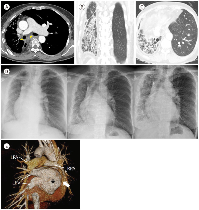
Unilateral pulmonary vein atresia (PVA) is a rare congenital cardiovascular anomaly occurring after the common pulmonary vein fails to incorporate into the left atrium. It is most commonly diagnosed in childhood, and diagnosis after reaching adulthood is extremely rare. Dyspnea on exertion and hemoptysis are common clinical features in adult PVA patients, whereas lung parenchymal abnormalities are indirect signs of PVA, which can manifest as interstitial lung disease. Herein, we present the case of a 62-year-old female diagnosed with unilateral PVA presenting as unilateral interstitial lung disease and report the changes in her chest radiographs over 12 years.

 求助内容:
求助内容: 应助结果提醒方式:


