Minji Shin, Young Jin Heo, Donghyun Kim, Hae Woong Jeong, Jin Wook Baek, Ha Young Park
{"title":"孤立的颅内模拟脑膜瘤的Rosai-Dorfman病1例报告。","authors":"Minji Shin, Young Jin Heo, Donghyun Kim, Hae Woong Jeong, Jin Wook Baek, Ha Young Park","doi":"10.3348/jksr.2021.0121","DOIUrl":null,"url":null,"abstract":"<p><p>Rosai-Dorfman Disease (RDD) is a rare lymphoproliferative disease, and the occurrence of isolated intracranial RDD is extremely rare. Most cases of intracranial RDDs present as dural masses showing homogenous enhancement on MRI, which makes it difficult to differentiate these masses from meningiomas before surgery unless massive cervical lymphadenopathy is observed. We herein report a rare case of isolated intracranial RDD in a 65-year-old male. Brain MRI revealed a well-defined enhancing mass-like lesion involving the right frontal convexity and subtle diffusion restriction. However, only a subtle blush was observed on the preoperative cerebral angiogram. Although instances of isolated intracranial RDD are rare, it should be considered as a potential differential diagnosis when a dural mass with hypovascularity is visualized on the cerebral angiogram.</p>","PeriodicalId":74904,"journal":{"name":"Taehan Yongsang Uihakhoe chi","volume":" ","pages":"719-723"},"PeriodicalIF":0.0000,"publicationDate":"2022-05-01","publicationTypes":"Journal Article","fieldsOfStudy":null,"isOpenAccess":false,"openAccessPdf":"https://ftp.ncbi.nlm.nih.gov/pub/pmc/oa_pdf/52/d4/jksr-83-719.PMC9514521.pdf","citationCount":"0","resultStr":"{\"title\":\"Isolated Intracranial Rosai-Dorfman Disease Mimicking Meningioma: A Case Report.\",\"authors\":\"Minji Shin, Young Jin Heo, Donghyun Kim, Hae Woong Jeong, Jin Wook Baek, Ha Young Park\",\"doi\":\"10.3348/jksr.2021.0121\",\"DOIUrl\":null,\"url\":null,\"abstract\":\"<p><p>Rosai-Dorfman Disease (RDD) is a rare lymphoproliferative disease, and the occurrence of isolated intracranial RDD is extremely rare. Most cases of intracranial RDDs present as dural masses showing homogenous enhancement on MRI, which makes it difficult to differentiate these masses from meningiomas before surgery unless massive cervical lymphadenopathy is observed. We herein report a rare case of isolated intracranial RDD in a 65-year-old male. Brain MRI revealed a well-defined enhancing mass-like lesion involving the right frontal convexity and subtle diffusion restriction. However, only a subtle blush was observed on the preoperative cerebral angiogram. Although instances of isolated intracranial RDD are rare, it should be considered as a potential differential diagnosis when a dural mass with hypovascularity is visualized on the cerebral angiogram.</p>\",\"PeriodicalId\":74904,\"journal\":{\"name\":\"Taehan Yongsang Uihakhoe chi\",\"volume\":\" \",\"pages\":\"719-723\"},\"PeriodicalIF\":0.0000,\"publicationDate\":\"2022-05-01\",\"publicationTypes\":\"Journal Article\",\"fieldsOfStudy\":null,\"isOpenAccess\":false,\"openAccessPdf\":\"https://ftp.ncbi.nlm.nih.gov/pub/pmc/oa_pdf/52/d4/jksr-83-719.PMC9514521.pdf\",\"citationCount\":\"0\",\"resultStr\":null,\"platform\":\"Semanticscholar\",\"paperid\":null,\"PeriodicalName\":\"Taehan Yongsang Uihakhoe chi\",\"FirstCategoryId\":\"1085\",\"ListUrlMain\":\"https://doi.org/10.3348/jksr.2021.0121\",\"RegionNum\":0,\"RegionCategory\":null,\"ArticlePicture\":[],\"TitleCN\":null,\"AbstractTextCN\":null,\"PMCID\":null,\"EPubDate\":\"2022/5/25 0:00:00\",\"PubModel\":\"Epub\",\"JCR\":\"\",\"JCRName\":\"\",\"Score\":null,\"Total\":0}","platform":"Semanticscholar","paperid":null,"PeriodicalName":"Taehan Yongsang Uihakhoe chi","FirstCategoryId":"1085","ListUrlMain":"https://doi.org/10.3348/jksr.2021.0121","RegionNum":0,"RegionCategory":null,"ArticlePicture":[],"TitleCN":null,"AbstractTextCN":null,"PMCID":null,"EPubDate":"2022/5/25 0:00:00","PubModel":"Epub","JCR":"","JCRName":"","Score":null,"Total":0}
Isolated Intracranial Rosai-Dorfman Disease Mimicking Meningioma: A Case Report.
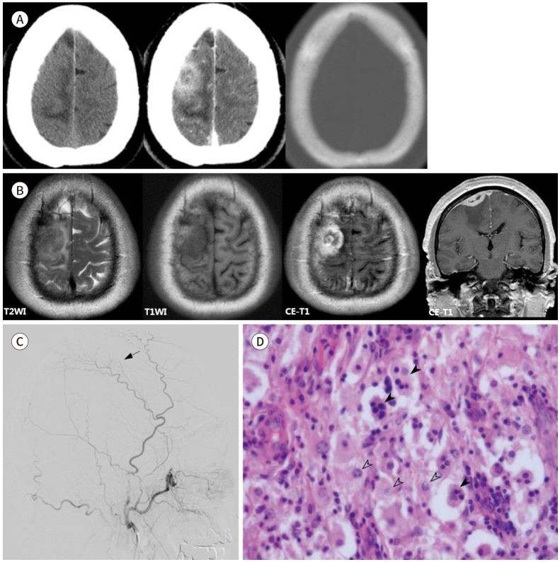
Rosai-Dorfman Disease (RDD) is a rare lymphoproliferative disease, and the occurrence of isolated intracranial RDD is extremely rare. Most cases of intracranial RDDs present as dural masses showing homogenous enhancement on MRI, which makes it difficult to differentiate these masses from meningiomas before surgery unless massive cervical lymphadenopathy is observed. We herein report a rare case of isolated intracranial RDD in a 65-year-old male. Brain MRI revealed a well-defined enhancing mass-like lesion involving the right frontal convexity and subtle diffusion restriction. However, only a subtle blush was observed on the preoperative cerebral angiogram. Although instances of isolated intracranial RDD are rare, it should be considered as a potential differential diagnosis when a dural mass with hypovascularity is visualized on the cerebral angiogram.

 求助内容:
求助内容: 应助结果提醒方式:


