Yosuke Hattori, Nobuyuki Asai, Shotaro Mori, Ken Ikuta, Yusuke Kazama, Yusuke Iesaki, Shimpei Takahashi, Atsushi Kaneko, Tomotaro Sato
{"title":"在日本接受全膝关节置换术的膝内翻骨性关节炎患者中,髓内矫正棒的股骨外翻矫正角度与股骨外侧弯曲密切相关。","authors":"Yosuke Hattori, Nobuyuki Asai, Shotaro Mori, Ken Ikuta, Yusuke Kazama, Yusuke Iesaki, Shimpei Takahashi, Atsushi Kaneko, Tomotaro Sato","doi":"10.1155/2022/7223534","DOIUrl":null,"url":null,"abstract":"<p><strong>Background: </strong>This study aimed to investigate factors, such as differences in femoral shape, that could affect the femoral valgus correction angle (VCA) for the intramedullary alignment rod (IM rod) by using a three-dimensional (3D) measurement system in patients with varus knee osteoarthritis undergoing total knee arthroplasty (TKA).</p><p><strong>Methods: </strong>A total of 305 knees in 233 Japanese patients with varus knee osteoarthritis who underwent primary TKA by using Jig Engaged 3D Pre-Operative Planning Software for the TKA operation support system was examined. We retrospectively analysed factors, such as the shape of the proximal, middle, and distal femur in the coronal plane, all of which could affect the VCA for the IM rod, by multiple linear regression analyses.</p><p><strong>Results: </strong>The VCA for the IM rod was 5.9° ± 1.6° (range: 1.7° to 10.7°), and the femoral lateral bowing angle (FBA) was 3.5° ± 3.2°. Major factors independently associated with the VCA for the IM rod were the FBA (<i>β</i>: 0.75), femoral offset (<i>β</i>: 0.38), and the medial angle between the mechanical femoral axis and the line that connects the distal margins of the medial and lateral femoral condyles (<i>β</i>: -0.16). The model was created by stepwise multiple linear regression (<i>F</i> = 266.6, <i>p</i> < 0.001, and estimated effect size = 4.4) explained 85% of the variance in the VCA for the IM rod (<i>R</i> <sup>2</sup> = 0.85).</p><p><strong>Conclusions: </strong>The VCA for the IM rod was most strongly associated with femoral lateral bowing in patients with varus knee osteoarthritis undergoing TKA. Our findings suggest that preoperatively measuring the VCA for the IM rod in patients with femoral lateral bowing by using a 3D measurement system could be useful for accurate coronal alignment of the femoral component in TKA.</p>","PeriodicalId":7358,"journal":{"name":"Advances in Orthopedics","volume":" ","pages":"7223534"},"PeriodicalIF":1.6000,"publicationDate":"2022-08-16","publicationTypes":"Journal Article","fieldsOfStudy":null,"isOpenAccess":false,"openAccessPdf":"https://www.ncbi.nlm.nih.gov/pmc/articles/PMC9398862/pdf/","citationCount":"1","resultStr":"{\"title\":\"Femoral Valgus Correction Angle for the Intramedullary Alignment Rod Is Strongly Associated with Femoral Lateral Bowing in Japanese Patients with Varus Knee Osteoarthritis Undergoing Total Knee Arthroplasty.\",\"authors\":\"Yosuke Hattori, Nobuyuki Asai, Shotaro Mori, Ken Ikuta, Yusuke Kazama, Yusuke Iesaki, Shimpei Takahashi, Atsushi Kaneko, Tomotaro Sato\",\"doi\":\"10.1155/2022/7223534\",\"DOIUrl\":null,\"url\":null,\"abstract\":\"<p><strong>Background: </strong>This study aimed to investigate factors, such as differences in femoral shape, that could affect the femoral valgus correction angle (VCA) for the intramedullary alignment rod (IM rod) by using a three-dimensional (3D) measurement system in patients with varus knee osteoarthritis undergoing total knee arthroplasty (TKA).</p><p><strong>Methods: </strong>A total of 305 knees in 233 Japanese patients with varus knee osteoarthritis who underwent primary TKA by using Jig Engaged 3D Pre-Operative Planning Software for the TKA operation support system was examined. We retrospectively analysed factors, such as the shape of the proximal, middle, and distal femur in the coronal plane, all of which could affect the VCA for the IM rod, by multiple linear regression analyses.</p><p><strong>Results: </strong>The VCA for the IM rod was 5.9° ± 1.6° (range: 1.7° to 10.7°), and the femoral lateral bowing angle (FBA) was 3.5° ± 3.2°. Major factors independently associated with the VCA for the IM rod were the FBA (<i>β</i>: 0.75), femoral offset (<i>β</i>: 0.38), and the medial angle between the mechanical femoral axis and the line that connects the distal margins of the medial and lateral femoral condyles (<i>β</i>: -0.16). The model was created by stepwise multiple linear regression (<i>F</i> = 266.6, <i>p</i> < 0.001, and estimated effect size = 4.4) explained 85% of the variance in the VCA for the IM rod (<i>R</i> <sup>2</sup> = 0.85).</p><p><strong>Conclusions: </strong>The VCA for the IM rod was most strongly associated with femoral lateral bowing in patients with varus knee osteoarthritis undergoing TKA. Our findings suggest that preoperatively measuring the VCA for the IM rod in patients with femoral lateral bowing by using a 3D measurement system could be useful for accurate coronal alignment of the femoral component in TKA.</p>\",\"PeriodicalId\":7358,\"journal\":{\"name\":\"Advances in Orthopedics\",\"volume\":\" \",\"pages\":\"7223534\"},\"PeriodicalIF\":1.6000,\"publicationDate\":\"2022-08-16\",\"publicationTypes\":\"Journal Article\",\"fieldsOfStudy\":null,\"isOpenAccess\":false,\"openAccessPdf\":\"https://www.ncbi.nlm.nih.gov/pmc/articles/PMC9398862/pdf/\",\"citationCount\":\"1\",\"resultStr\":null,\"platform\":\"Semanticscholar\",\"paperid\":null,\"PeriodicalName\":\"Advances in Orthopedics\",\"FirstCategoryId\":\"1085\",\"ListUrlMain\":\"https://doi.org/10.1155/2022/7223534\",\"RegionNum\":0,\"RegionCategory\":null,\"ArticlePicture\":[],\"TitleCN\":null,\"AbstractTextCN\":null,\"PMCID\":null,\"EPubDate\":\"2022/1/1 0:00:00\",\"PubModel\":\"eCollection\",\"JCR\":\"Q3\",\"JCRName\":\"ORTHOPEDICS\",\"Score\":null,\"Total\":0}","platform":"Semanticscholar","paperid":null,"PeriodicalName":"Advances in Orthopedics","FirstCategoryId":"1085","ListUrlMain":"https://doi.org/10.1155/2022/7223534","RegionNum":0,"RegionCategory":null,"ArticlePicture":[],"TitleCN":null,"AbstractTextCN":null,"PMCID":null,"EPubDate":"2022/1/1 0:00:00","PubModel":"eCollection","JCR":"Q3","JCRName":"ORTHOPEDICS","Score":null,"Total":0}
Femoral Valgus Correction Angle for the Intramedullary Alignment Rod Is Strongly Associated with Femoral Lateral Bowing in Japanese Patients with Varus Knee Osteoarthritis Undergoing Total Knee Arthroplasty.
Background: This study aimed to investigate factors, such as differences in femoral shape, that could affect the femoral valgus correction angle (VCA) for the intramedullary alignment rod (IM rod) by using a three-dimensional (3D) measurement system in patients with varus knee osteoarthritis undergoing total knee arthroplasty (TKA).
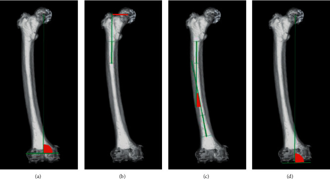
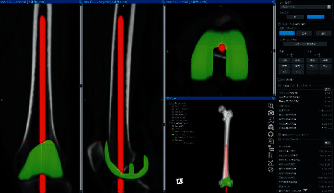
Methods: A total of 305 knees in 233 Japanese patients with varus knee osteoarthritis who underwent primary TKA by using Jig Engaged 3D Pre-Operative Planning Software for the TKA operation support system was examined. We retrospectively analysed factors, such as the shape of the proximal, middle, and distal femur in the coronal plane, all of which could affect the VCA for the IM rod, by multiple linear regression analyses.
Results: The VCA for the IM rod was 5.9° ± 1.6° (range: 1.7° to 10.7°), and the femoral lateral bowing angle (FBA) was 3.5° ± 3.2°. Major factors independently associated with the VCA for the IM rod were the FBA (β: 0.75), femoral offset (β: 0.38), and the medial angle between the mechanical femoral axis and the line that connects the distal margins of the medial and lateral femoral condyles (β: -0.16). The model was created by stepwise multiple linear regression (F = 266.6, p < 0.001, and estimated effect size = 4.4) explained 85% of the variance in the VCA for the IM rod (R2 = 0.85).
Conclusions: The VCA for the IM rod was most strongly associated with femoral lateral bowing in patients with varus knee osteoarthritis undergoing TKA. Our findings suggest that preoperatively measuring the VCA for the IM rod in patients with femoral lateral bowing by using a 3D measurement system could be useful for accurate coronal alignment of the femoral component in TKA.
期刊介绍:
Advances in Orthopedics is a peer-reviewed, Open Access journal that provides a forum for orthopaedics working on improving the quality of orthopedic health care. The journal publishes original research articles, review articles, and clinical studies related to arthroplasty, hand surgery, limb reconstruction, pediatric orthopaedics, sports medicine, trauma, spinal deformities, and orthopaedic oncology.

 求助内容:
求助内容: 应助结果提醒方式:


