{"title":"微管的边缘带在视网膜双极突触末端运输和组织线粒体。","authors":"Malkolm Graffe, David Zenisek, Justin W Taraska","doi":"10.1085/jgp.201511396","DOIUrl":null,"url":null,"abstract":"<p><p>A set of bipolar cells in the retina of goldfish contains giant synaptic terminals that can be over 10 µm in diameter. Hundreds of thousands of synaptic vesicles fill these terminals and engage in continuous rounds of exocytosis. How the cytoskeleton and other organelles in these neurons are organized to control synaptic activity is unknown. Here, we used 3-D fluorescence and 3-D electron microscopy to visualize the complex subcellular architecture of these terminals. We discovered a thick band of microtubules that emerged from the axon to loop around the terminal periphery throughout the presynaptic space. This previously unknown microtubule structure associated with a substantial population of mitochondria in the synaptic terminal. Drugs that inhibit microtubule-based kinesin motors led to accumulation of mitochondria in the axon. We conclude that this prominent microtubule band is crucial to the transport and localization of mitochondria into the presynaptic space to provide the sustained energy necessary for continuous transmitter release in these giant synaptic terminals. </p>","PeriodicalId":173753,"journal":{"name":"The Journal of General Physiology","volume":" ","pages":"109-17"},"PeriodicalIF":0.0000,"publicationDate":"2015-07-01","publicationTypes":"Journal Article","fieldsOfStudy":null,"isOpenAccess":false,"openAccessPdf":"https://sci-hub-pdf.com/10.1085/jgp.201511396","citationCount":"24","resultStr":"{\"title\":\"A marginal band of microtubules transports and organizes mitochondria in retinal bipolar synaptic terminals.\",\"authors\":\"Malkolm Graffe, David Zenisek, Justin W Taraska\",\"doi\":\"10.1085/jgp.201511396\",\"DOIUrl\":null,\"url\":null,\"abstract\":\"<p><p>A set of bipolar cells in the retina of goldfish contains giant synaptic terminals that can be over 10 µm in diameter. Hundreds of thousands of synaptic vesicles fill these terminals and engage in continuous rounds of exocytosis. How the cytoskeleton and other organelles in these neurons are organized to control synaptic activity is unknown. Here, we used 3-D fluorescence and 3-D electron microscopy to visualize the complex subcellular architecture of these terminals. We discovered a thick band of microtubules that emerged from the axon to loop around the terminal periphery throughout the presynaptic space. This previously unknown microtubule structure associated with a substantial population of mitochondria in the synaptic terminal. Drugs that inhibit microtubule-based kinesin motors led to accumulation of mitochondria in the axon. We conclude that this prominent microtubule band is crucial to the transport and localization of mitochondria into the presynaptic space to provide the sustained energy necessary for continuous transmitter release in these giant synaptic terminals. </p>\",\"PeriodicalId\":173753,\"journal\":{\"name\":\"The Journal of General Physiology\",\"volume\":\" \",\"pages\":\"109-17\"},\"PeriodicalIF\":0.0000,\"publicationDate\":\"2015-07-01\",\"publicationTypes\":\"Journal Article\",\"fieldsOfStudy\":null,\"isOpenAccess\":false,\"openAccessPdf\":\"https://sci-hub-pdf.com/10.1085/jgp.201511396\",\"citationCount\":\"24\",\"resultStr\":null,\"platform\":\"Semanticscholar\",\"paperid\":null,\"PeriodicalName\":\"The Journal of General Physiology\",\"FirstCategoryId\":\"3\",\"ListUrlMain\":\"https://doi.org/10.1085/jgp.201511396\",\"RegionNum\":0,\"RegionCategory\":null,\"ArticlePicture\":[],\"TitleCN\":null,\"AbstractTextCN\":null,\"PMCID\":null,\"EPubDate\":\"\",\"PubModel\":\"\",\"JCR\":\"\",\"JCRName\":\"\",\"Score\":null,\"Total\":0}","platform":"Semanticscholar","paperid":null,"PeriodicalName":"The Journal of General Physiology","FirstCategoryId":"3","ListUrlMain":"https://doi.org/10.1085/jgp.201511396","RegionNum":0,"RegionCategory":null,"ArticlePicture":[],"TitleCN":null,"AbstractTextCN":null,"PMCID":null,"EPubDate":"","PubModel":"","JCR":"","JCRName":"","Score":null,"Total":0}
A marginal band of microtubules transports and organizes mitochondria in retinal bipolar synaptic terminals.
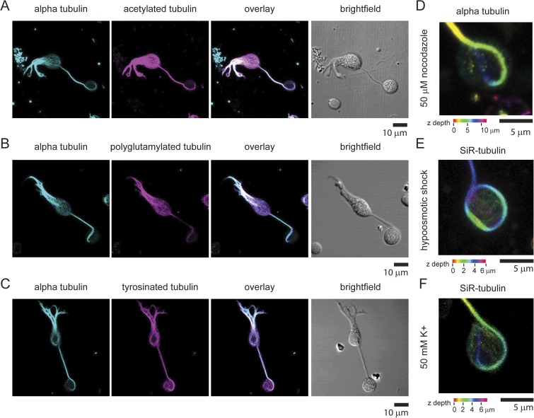
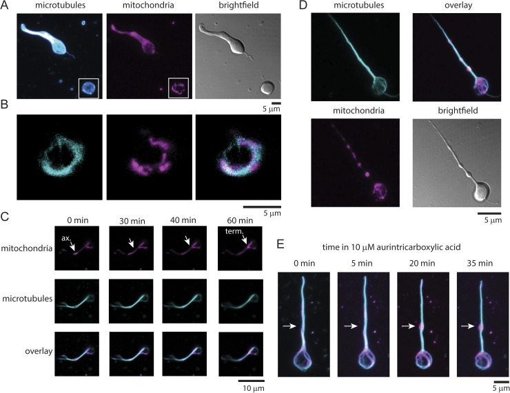
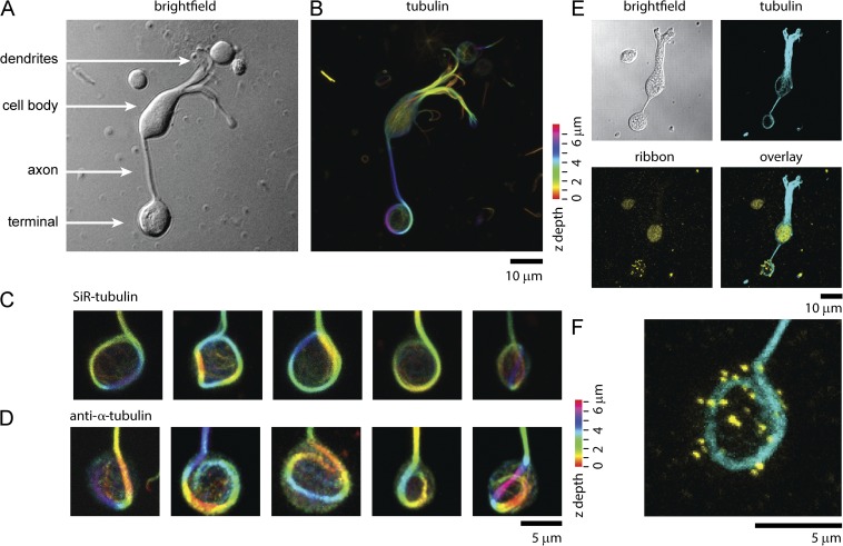
A set of bipolar cells in the retina of goldfish contains giant synaptic terminals that can be over 10 µm in diameter. Hundreds of thousands of synaptic vesicles fill these terminals and engage in continuous rounds of exocytosis. How the cytoskeleton and other organelles in these neurons are organized to control synaptic activity is unknown. Here, we used 3-D fluorescence and 3-D electron microscopy to visualize the complex subcellular architecture of these terminals. We discovered a thick band of microtubules that emerged from the axon to loop around the terminal periphery throughout the presynaptic space. This previously unknown microtubule structure associated with a substantial population of mitochondria in the synaptic terminal. Drugs that inhibit microtubule-based kinesin motors led to accumulation of mitochondria in the axon. We conclude that this prominent microtubule band is crucial to the transport and localization of mitochondria into the presynaptic space to provide the sustained energy necessary for continuous transmitter release in these giant synaptic terminals.

 求助内容:
求助内容: 应助结果提醒方式:


