Jae Heon Kim, Hong Jun Lee, Seung Hwan Doo, Won Jae Yang, Dongho Choi, Jung Hoon Kim, Jong Ho Won, Yun Seob Song
{"title":"应用纳米颗粒监测人间充质干细胞移植到勃起功能障碍大鼠阴茎海绵体的效果。","authors":"Jae Heon Kim, Hong Jun Lee, Seung Hwan Doo, Won Jae Yang, Dongho Choi, Jung Hoon Kim, Jong Ho Won, Yun Seob Song","doi":"10.4111/kju.2015.56.4.280","DOIUrl":null,"url":null,"abstract":"<p><strong>Purpose: </strong>This study was performed to examine the treatment of erectile dysfunction by use of superparamagnetic iron oxide nanoparticles-labeled human mesenchymal stem cells (SPION-MSCs) transplanted into the cavernous nerve injured cavernosa of rats as monitored by molecular magnetic resonance imaging (MRI).</p><p><strong>Materials and methods: </strong>Eight-week-old male Sprague-Dawley rats were divided into three groups of 10 rats each: group 1, sham operation; group 2, cavernous nerve injury; group 3, SPION-MSC treatment after cavernous nerve injury. Immediately after the cavernous nerve injury in group 3, SPION-MSCs were injected into the cavernous nerve injured cavernosa. Serial T2-weighted MRI was done immediately after injection and at 2 and 4 weeks. Erectile response was assessed by cavernous nerve stimulation at 2 and 4 weeks.</p><p><strong>Results: </strong>Prussian blue staining of SPION-MSCs revealed abundant uptake of SPION in the cytoplasm. After injection of 1×10(6) SPION-MSCs into the cavernosa of rats, T2-weighted MRI showed a clear hypointense signal induced by the injection. The presence of SPION in the corpora cavernosa was confirmed with Prussian blue staining. At 2 and 4 weeks, rats with cavernous nerve injury had significantly lower erectile function than did rats without cavernous nerve injury (p<0.05). The group transplanted with SPION-MSCs showed higher erectile function than did the group without SPION-MSCs (p<0.05). The presence of SPION-MSCs for up to 4 weeks was confirmed by MRI imaging and Prussian blue staining in the corpus cavernosa.</p><p><strong>Conclusions: </strong>Transplanted SPION-MSCs existed for up to 4 weeks in the cavernous nerve injured cavernosa of rats. Erectile dysfunction recovered and could be monitored by MRI.</p>","PeriodicalId":17819,"journal":{"name":"Korean Journal of Urology","volume":"56 4","pages":"280-7"},"PeriodicalIF":0.0000,"publicationDate":"2015-04-01","publicationTypes":"Journal Article","fieldsOfStudy":null,"isOpenAccess":false,"openAccessPdf":"https://sci-hub-pdf.com/10.4111/kju.2015.56.4.280","citationCount":"12","resultStr":"{\"title\":\"Use of nanoparticles to monitor human mesenchymal stem cells transplanted into penile cavernosum of rats with erectile dysfunction.\",\"authors\":\"Jae Heon Kim, Hong Jun Lee, Seung Hwan Doo, Won Jae Yang, Dongho Choi, Jung Hoon Kim, Jong Ho Won, Yun Seob Song\",\"doi\":\"10.4111/kju.2015.56.4.280\",\"DOIUrl\":null,\"url\":null,\"abstract\":\"<p><strong>Purpose: </strong>This study was performed to examine the treatment of erectile dysfunction by use of superparamagnetic iron oxide nanoparticles-labeled human mesenchymal stem cells (SPION-MSCs) transplanted into the cavernous nerve injured cavernosa of rats as monitored by molecular magnetic resonance imaging (MRI).</p><p><strong>Materials and methods: </strong>Eight-week-old male Sprague-Dawley rats were divided into three groups of 10 rats each: group 1, sham operation; group 2, cavernous nerve injury; group 3, SPION-MSC treatment after cavernous nerve injury. Immediately after the cavernous nerve injury in group 3, SPION-MSCs were injected into the cavernous nerve injured cavernosa. Serial T2-weighted MRI was done immediately after injection and at 2 and 4 weeks. Erectile response was assessed by cavernous nerve stimulation at 2 and 4 weeks.</p><p><strong>Results: </strong>Prussian blue staining of SPION-MSCs revealed abundant uptake of SPION in the cytoplasm. After injection of 1×10(6) SPION-MSCs into the cavernosa of rats, T2-weighted MRI showed a clear hypointense signal induced by the injection. The presence of SPION in the corpora cavernosa was confirmed with Prussian blue staining. At 2 and 4 weeks, rats with cavernous nerve injury had significantly lower erectile function than did rats without cavernous nerve injury (p<0.05). The group transplanted with SPION-MSCs showed higher erectile function than did the group without SPION-MSCs (p<0.05). The presence of SPION-MSCs for up to 4 weeks was confirmed by MRI imaging and Prussian blue staining in the corpus cavernosa.</p><p><strong>Conclusions: </strong>Transplanted SPION-MSCs existed for up to 4 weeks in the cavernous nerve injured cavernosa of rats. Erectile dysfunction recovered and could be monitored by MRI.</p>\",\"PeriodicalId\":17819,\"journal\":{\"name\":\"Korean Journal of Urology\",\"volume\":\"56 4\",\"pages\":\"280-7\"},\"PeriodicalIF\":0.0000,\"publicationDate\":\"2015-04-01\",\"publicationTypes\":\"Journal Article\",\"fieldsOfStudy\":null,\"isOpenAccess\":false,\"openAccessPdf\":\"https://sci-hub-pdf.com/10.4111/kju.2015.56.4.280\",\"citationCount\":\"12\",\"resultStr\":null,\"platform\":\"Semanticscholar\",\"paperid\":null,\"PeriodicalName\":\"Korean Journal of Urology\",\"FirstCategoryId\":\"1085\",\"ListUrlMain\":\"https://doi.org/10.4111/kju.2015.56.4.280\",\"RegionNum\":0,\"RegionCategory\":null,\"ArticlePicture\":[],\"TitleCN\":null,\"AbstractTextCN\":null,\"PMCID\":null,\"EPubDate\":\"2015/3/20 0:00:00\",\"PubModel\":\"Epub\",\"JCR\":\"\",\"JCRName\":\"\",\"Score\":null,\"Total\":0}","platform":"Semanticscholar","paperid":null,"PeriodicalName":"Korean Journal of Urology","FirstCategoryId":"1085","ListUrlMain":"https://doi.org/10.4111/kju.2015.56.4.280","RegionNum":0,"RegionCategory":null,"ArticlePicture":[],"TitleCN":null,"AbstractTextCN":null,"PMCID":null,"EPubDate":"2015/3/20 0:00:00","PubModel":"Epub","JCR":"","JCRName":"","Score":null,"Total":0}
Use of nanoparticles to monitor human mesenchymal stem cells transplanted into penile cavernosum of rats with erectile dysfunction.
Purpose: This study was performed to examine the treatment of erectile dysfunction by use of superparamagnetic iron oxide nanoparticles-labeled human mesenchymal stem cells (SPION-MSCs) transplanted into the cavernous nerve injured cavernosa of rats as monitored by molecular magnetic resonance imaging (MRI).
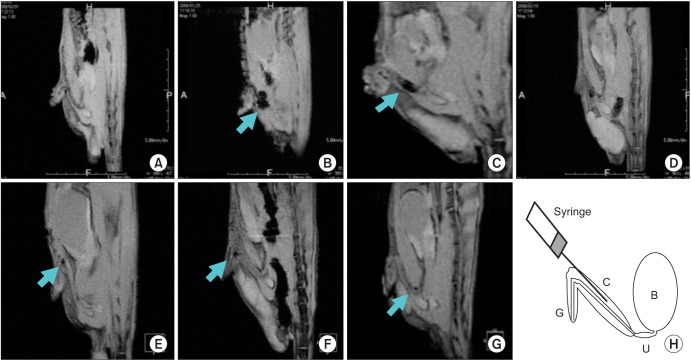
Materials and methods: Eight-week-old male Sprague-Dawley rats were divided into three groups of 10 rats each: group 1, sham operation; group 2, cavernous nerve injury; group 3, SPION-MSC treatment after cavernous nerve injury. Immediately after the cavernous nerve injury in group 3, SPION-MSCs were injected into the cavernous nerve injured cavernosa. Serial T2-weighted MRI was done immediately after injection and at 2 and 4 weeks. Erectile response was assessed by cavernous nerve stimulation at 2 and 4 weeks.
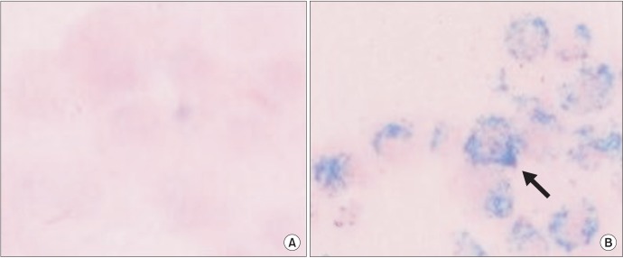
Results: Prussian blue staining of SPION-MSCs revealed abundant uptake of SPION in the cytoplasm. After injection of 1×10(6) SPION-MSCs into the cavernosa of rats, T2-weighted MRI showed a clear hypointense signal induced by the injection. The presence of SPION in the corpora cavernosa was confirmed with Prussian blue staining. At 2 and 4 weeks, rats with cavernous nerve injury had significantly lower erectile function than did rats without cavernous nerve injury (p<0.05). The group transplanted with SPION-MSCs showed higher erectile function than did the group without SPION-MSCs (p<0.05). The presence of SPION-MSCs for up to 4 weeks was confirmed by MRI imaging and Prussian blue staining in the corpus cavernosa.
Conclusions: Transplanted SPION-MSCs existed for up to 4 weeks in the cavernous nerve injured cavernosa of rats. Erectile dysfunction recovered and could be monitored by MRI.

 求助内容:
求助内容: 应助结果提醒方式:


