Ümit Naykı, Cenk Naykı, Paşa Uluğ, Ismayil Yılmaz, Zeliha Cetin, Yusuf Yıldırım
{"title":"一例罕见的巨大囊性平滑肌瘤表现为腹膜后肿块。","authors":"Ümit Naykı, Cenk Naykı, Paşa Uluğ, Ismayil Yılmaz, Zeliha Cetin, Yusuf Yıldırım","doi":"","DOIUrl":null,"url":null,"abstract":"<p><strong>Background: </strong>Giant retroperitoneal uterine leiomyomas are uncommon. Degenerative changes of a leiomyoma may lead to unusual presentation resulting in misdiagnosis preoperatively. The final diagnosis can be made either intraoperatively or histologically.</p><p><strong>Case: </strong>We report a 45-year-old multiparous women presented with abdominal distension and fatigue for six months. Abdominopelvic Sonography and computed tomography showed a large cystic mass that filled the pelvis and abdomen. With the preoperative diagnosis of a malignant tumor, a laparotomy was planned. Intraoperatively, a cystic mass originated from the uterus near the left side of the broad ligament extending to the retroperitoneal space was observed. Total hysterectomy and bilateral salphingo-oopherectomy was administered. The histology revealed a leiomyoma with cystic degeneration.</p><p><strong>Conclusion: </strong>Retroperitoneal leiomyomas should be kept in mind in the diferrential diagnosis of a giant cystic mass in abdomen.</p>","PeriodicalId":14673,"journal":{"name":"Iranian Journal of Reproductive Medicine","volume":"12 12","pages":"831-4"},"PeriodicalIF":0.0000,"publicationDate":"2014-12-01","publicationTypes":"Journal Article","fieldsOfStudy":null,"isOpenAccess":false,"openAccessPdf":"https://www.ncbi.nlm.nih.gov/pmc/articles/PMC4330664/pdf/","citationCount":"0","resultStr":"{\"title\":\"A rare case of a giant cystic leiomyoma presenting as a retroperitoneal mass.\",\"authors\":\"Ümit Naykı, Cenk Naykı, Paşa Uluğ, Ismayil Yılmaz, Zeliha Cetin, Yusuf Yıldırım\",\"doi\":\"\",\"DOIUrl\":null,\"url\":null,\"abstract\":\"<p><strong>Background: </strong>Giant retroperitoneal uterine leiomyomas are uncommon. Degenerative changes of a leiomyoma may lead to unusual presentation resulting in misdiagnosis preoperatively. The final diagnosis can be made either intraoperatively or histologically.</p><p><strong>Case: </strong>We report a 45-year-old multiparous women presented with abdominal distension and fatigue for six months. Abdominopelvic Sonography and computed tomography showed a large cystic mass that filled the pelvis and abdomen. With the preoperative diagnosis of a malignant tumor, a laparotomy was planned. Intraoperatively, a cystic mass originated from the uterus near the left side of the broad ligament extending to the retroperitoneal space was observed. Total hysterectomy and bilateral salphingo-oopherectomy was administered. The histology revealed a leiomyoma with cystic degeneration.</p><p><strong>Conclusion: </strong>Retroperitoneal leiomyomas should be kept in mind in the diferrential diagnosis of a giant cystic mass in abdomen.</p>\",\"PeriodicalId\":14673,\"journal\":{\"name\":\"Iranian Journal of Reproductive Medicine\",\"volume\":\"12 12\",\"pages\":\"831-4\"},\"PeriodicalIF\":0.0000,\"publicationDate\":\"2014-12-01\",\"publicationTypes\":\"Journal Article\",\"fieldsOfStudy\":null,\"isOpenAccess\":false,\"openAccessPdf\":\"https://www.ncbi.nlm.nih.gov/pmc/articles/PMC4330664/pdf/\",\"citationCount\":\"0\",\"resultStr\":null,\"platform\":\"Semanticscholar\",\"paperid\":null,\"PeriodicalName\":\"Iranian Journal of Reproductive Medicine\",\"FirstCategoryId\":\"1085\",\"ListUrlMain\":\"\",\"RegionNum\":0,\"RegionCategory\":null,\"ArticlePicture\":[],\"TitleCN\":null,\"AbstractTextCN\":null,\"PMCID\":null,\"EPubDate\":\"\",\"PubModel\":\"\",\"JCR\":\"\",\"JCRName\":\"\",\"Score\":null,\"Total\":0}","platform":"Semanticscholar","paperid":null,"PeriodicalName":"Iranian Journal of Reproductive Medicine","FirstCategoryId":"1085","ListUrlMain":"","RegionNum":0,"RegionCategory":null,"ArticlePicture":[],"TitleCN":null,"AbstractTextCN":null,"PMCID":null,"EPubDate":"","PubModel":"","JCR":"","JCRName":"","Score":null,"Total":0}
A rare case of a giant cystic leiomyoma presenting as a retroperitoneal mass.
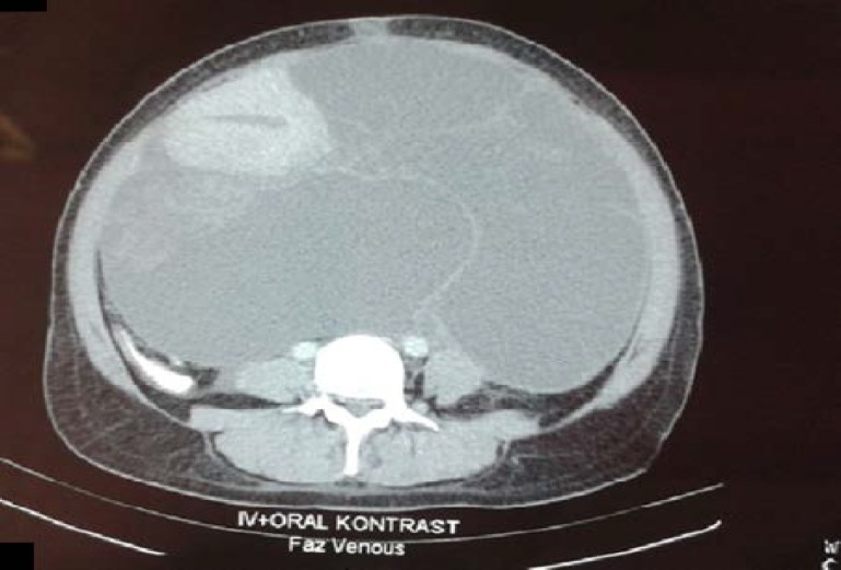
Background: Giant retroperitoneal uterine leiomyomas are uncommon. Degenerative changes of a leiomyoma may lead to unusual presentation resulting in misdiagnosis preoperatively. The final diagnosis can be made either intraoperatively or histologically.
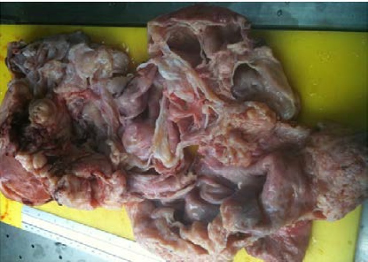
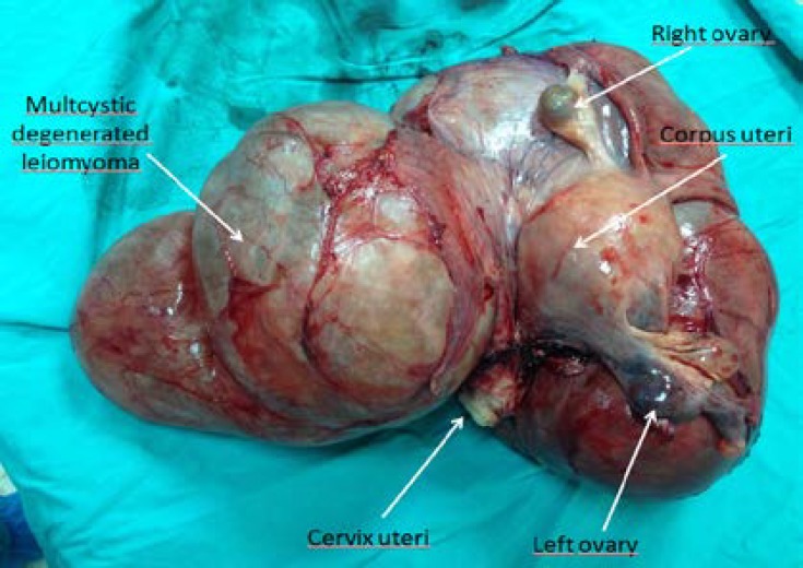
Case: We report a 45-year-old multiparous women presented with abdominal distension and fatigue for six months. Abdominopelvic Sonography and computed tomography showed a large cystic mass that filled the pelvis and abdomen. With the preoperative diagnosis of a malignant tumor, a laparotomy was planned. Intraoperatively, a cystic mass originated from the uterus near the left side of the broad ligament extending to the retroperitoneal space was observed. Total hysterectomy and bilateral salphingo-oopherectomy was administered. The histology revealed a leiomyoma with cystic degeneration.
Conclusion: Retroperitoneal leiomyomas should be kept in mind in the diferrential diagnosis of a giant cystic mass in abdomen.
 求助内容:
求助内容: 应助结果提醒方式:


