Danielle L Ippolito, John A Lewis, Chenggang Yu, Lisa R Leon, Jonathan D Stallings
{"title":"清醒大鼠热应激期间和之后循环代谢物的改变:暴露和器官特异性损伤的潜在生物标志物。","authors":"Danielle L Ippolito, John A Lewis, Chenggang Yu, Lisa R Leon, Jonathan D Stallings","doi":"10.1186/s12899-014-0014-0","DOIUrl":null,"url":null,"abstract":"<p><strong>Background: </strong>Heat illness is a debilitating and potentially life-threatening condition. Limited data are available to identify individuals with heat illness at greatest risk for organ damage. We recently described the transcriptomic and proteomic responses to heat injury and recovery in multiple organs in an in vivo model of conscious rats heated to a maximum core temperature of 41.8°C (Tc,Max). In this study, we examined changes in plasma metabolic networks at Tc,Max, 24, or 48 hours after the heat stress stimulus.</p><p><strong>Results: </strong>Circulating metabolites were identified by gas chromatography/mass spectrometry and liquid chromatography/tandem mass spectrometry. Bioinformatics analysis of the metabolomic data corroborated proteomics and transcriptomics data in the tissue at the pathway level, supporting modulations in metabolic networks including cell death or catabolism (pyrimidine and purine degradation, acetylation, sulfation, redox alterations and glutathione metabolism, and the urea cycle/creatinine metabolism), energetics (stasis in glycolysis and tricarboxylic acid cycle, β-oxidation), cholesterol and nitric oxide metabolism, and bile acids. Hierarchical clustering identified 15 biochemicals that differentiated animals with histopathological evidence of cardiac injury at 48 hours from uninjured animals. The metabolic networks perturbed in the plasma corroborated the tissue proteomics and transcriptomics pathway data, supporting a model of irreversible cell death and decrements in energetics as key indicators of cardiac damage in response to heat stress.</p><p><strong>Conclusions: </strong>Integrating plasma metabolomics with tissue proteomics and transcriptomics supports a diagnostic approach to assessing individual susceptibility to organ injury and predicting recovery after heat stress.</p>","PeriodicalId":35905,"journal":{"name":"BMC Physiology","volume":"14 ","pages":"14"},"PeriodicalIF":0.0000,"publicationDate":"2014-12-24","publicationTypes":"Journal Article","fieldsOfStudy":null,"isOpenAccess":false,"openAccessPdf":"https://sci-hub-pdf.com/10.1186/s12899-014-0014-0","citationCount":"53","resultStr":"{\"title\":\"Alteration in circulating metabolites during and after heat stress in the conscious rat: potential biomarkers of exposure and organ-specific injury.\",\"authors\":\"Danielle L Ippolito, John A Lewis, Chenggang Yu, Lisa R Leon, Jonathan D Stallings\",\"doi\":\"10.1186/s12899-014-0014-0\",\"DOIUrl\":null,\"url\":null,\"abstract\":\"<p><strong>Background: </strong>Heat illness is a debilitating and potentially life-threatening condition. Limited data are available to identify individuals with heat illness at greatest risk for organ damage. We recently described the transcriptomic and proteomic responses to heat injury and recovery in multiple organs in an in vivo model of conscious rats heated to a maximum core temperature of 41.8°C (Tc,Max). In this study, we examined changes in plasma metabolic networks at Tc,Max, 24, or 48 hours after the heat stress stimulus.</p><p><strong>Results: </strong>Circulating metabolites were identified by gas chromatography/mass spectrometry and liquid chromatography/tandem mass spectrometry. Bioinformatics analysis of the metabolomic data corroborated proteomics and transcriptomics data in the tissue at the pathway level, supporting modulations in metabolic networks including cell death or catabolism (pyrimidine and purine degradation, acetylation, sulfation, redox alterations and glutathione metabolism, and the urea cycle/creatinine metabolism), energetics (stasis in glycolysis and tricarboxylic acid cycle, β-oxidation), cholesterol and nitric oxide metabolism, and bile acids. Hierarchical clustering identified 15 biochemicals that differentiated animals with histopathological evidence of cardiac injury at 48 hours from uninjured animals. The metabolic networks perturbed in the plasma corroborated the tissue proteomics and transcriptomics pathway data, supporting a model of irreversible cell death and decrements in energetics as key indicators of cardiac damage in response to heat stress.</p><p><strong>Conclusions: </strong>Integrating plasma metabolomics with tissue proteomics and transcriptomics supports a diagnostic approach to assessing individual susceptibility to organ injury and predicting recovery after heat stress.</p>\",\"PeriodicalId\":35905,\"journal\":{\"name\":\"BMC Physiology\",\"volume\":\"14 \",\"pages\":\"14\"},\"PeriodicalIF\":0.0000,\"publicationDate\":\"2014-12-24\",\"publicationTypes\":\"Journal Article\",\"fieldsOfStudy\":null,\"isOpenAccess\":false,\"openAccessPdf\":\"https://sci-hub-pdf.com/10.1186/s12899-014-0014-0\",\"citationCount\":\"53\",\"resultStr\":null,\"platform\":\"Semanticscholar\",\"paperid\":null,\"PeriodicalName\":\"BMC Physiology\",\"FirstCategoryId\":\"1085\",\"ListUrlMain\":\"https://doi.org/10.1186/s12899-014-0014-0\",\"RegionNum\":0,\"RegionCategory\":null,\"ArticlePicture\":[],\"TitleCN\":null,\"AbstractTextCN\":null,\"PMCID\":null,\"EPubDate\":\"\",\"PubModel\":\"\",\"JCR\":\"Q1\",\"JCRName\":\"Biochemistry, Genetics and Molecular Biology\",\"Score\":null,\"Total\":0}","platform":"Semanticscholar","paperid":null,"PeriodicalName":"BMC Physiology","FirstCategoryId":"1085","ListUrlMain":"https://doi.org/10.1186/s12899-014-0014-0","RegionNum":0,"RegionCategory":null,"ArticlePicture":[],"TitleCN":null,"AbstractTextCN":null,"PMCID":null,"EPubDate":"","PubModel":"","JCR":"Q1","JCRName":"Biochemistry, Genetics and Molecular Biology","Score":null,"Total":0}
Alteration in circulating metabolites during and after heat stress in the conscious rat: potential biomarkers of exposure and organ-specific injury.
Background: Heat illness is a debilitating and potentially life-threatening condition. Limited data are available to identify individuals with heat illness at greatest risk for organ damage. We recently described the transcriptomic and proteomic responses to heat injury and recovery in multiple organs in an in vivo model of conscious rats heated to a maximum core temperature of 41.8°C (Tc,Max). In this study, we examined changes in plasma metabolic networks at Tc,Max, 24, or 48 hours after the heat stress stimulus.
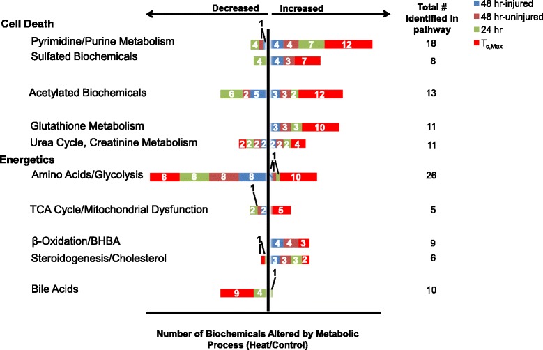
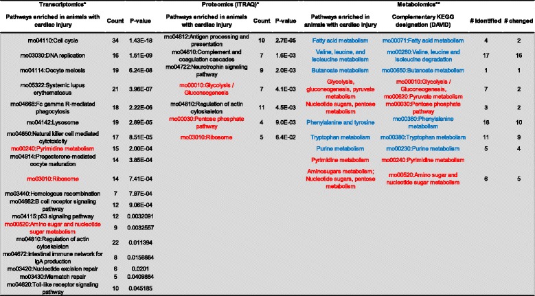
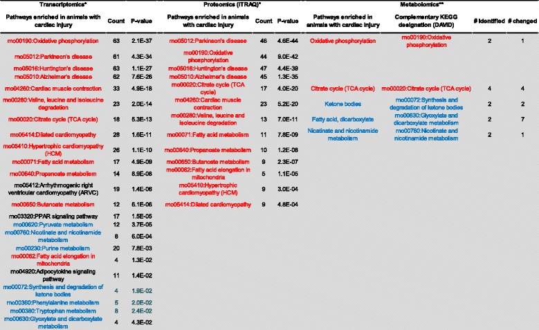
Results: Circulating metabolites were identified by gas chromatography/mass spectrometry and liquid chromatography/tandem mass spectrometry. Bioinformatics analysis of the metabolomic data corroborated proteomics and transcriptomics data in the tissue at the pathway level, supporting modulations in metabolic networks including cell death or catabolism (pyrimidine and purine degradation, acetylation, sulfation, redox alterations and glutathione metabolism, and the urea cycle/creatinine metabolism), energetics (stasis in glycolysis and tricarboxylic acid cycle, β-oxidation), cholesterol and nitric oxide metabolism, and bile acids. Hierarchical clustering identified 15 biochemicals that differentiated animals with histopathological evidence of cardiac injury at 48 hours from uninjured animals. The metabolic networks perturbed in the plasma corroborated the tissue proteomics and transcriptomics pathway data, supporting a model of irreversible cell death and decrements in energetics as key indicators of cardiac damage in response to heat stress.
Conclusions: Integrating plasma metabolomics with tissue proteomics and transcriptomics supports a diagnostic approach to assessing individual susceptibility to organ injury and predicting recovery after heat stress.
BMC PhysiologyBiochemistry, Genetics and Molecular Biology-Physiology
CiteScore
9.60
自引率
0.00%
发文量
0
期刊介绍:
BMC Physiology is an open access journal publishing original peer-reviewed research articles in cellular, tissue-level, organismal, functional, and developmental aspects of physiological processes. BMC Physiology (ISSN 1472-6793) is indexed/tracked/covered by PubMed, MEDLINE, BIOSIS, CAS, EMBASE, Scopus, Zoological Record and Google Scholar.

 求助内容:
求助内容: 应助结果提醒方式:


