Faten A Dawood, Rahmita W Rahmat, Suhaini B Kadiman, Lili N Abdullah, Mohd D Zamrin
{"title":"三维超声心动图数据集4片右心室心内膜轮廓提取的混合方法。","authors":"Faten A Dawood, Rahmita W Rahmat, Suhaini B Kadiman, Lili N Abdullah, Mohd D Zamrin","doi":"10.1155/2014/207149","DOIUrl":null,"url":null,"abstract":"<p><p>This paper presents a hybrid method to extract endocardial contour of the right ventricular (RV) in 4-slices from 3D echocardiography dataset. The overall framework comprises four processing phases. In Phase I, the region of interest (ROI) is identified by estimating the cavity boundary. Speckle noise reduction and contrast enhancement were implemented in Phase II as preprocessing tasks. In Phase III, the RV cavity region was segmented by generating intensity threshold which was used for once for all frames. Finally, Phase IV is proposed to extract the RV endocardial contour in a complete cardiac cycle using a combination of shape-based contour detection and improved radial search algorithm. The proposed method was applied to 16 datasets of 3D echocardiography encompassing the RV in long-axis view. The accuracy of experimental results obtained by the proposed method was evaluated qualitatively and quantitatively. It has been done by comparing the segmentation results of RV cavity based on endocardial contour extraction with the ground truth. The comparative analysis results show that the proposed method performs efficiently in all datasets with overall performance of 95% and the root mean square distances (RMSD) measure in terms of mean ± SD was found to be 2.21 ± 0.35 mm for RV endocardial contours. </p>","PeriodicalId":39059,"journal":{"name":"Advances in Bioinformatics","volume":"2014 ","pages":"207149"},"PeriodicalIF":0.0000,"publicationDate":"2014-01-01","publicationTypes":"Journal Article","fieldsOfStudy":null,"isOpenAccess":false,"openAccessPdf":"https://sci-hub-pdf.com/10.1155/2014/207149","citationCount":"1","resultStr":"{\"title\":\"A Hybrid Method for Endocardial Contour Extraction of Right Ventricle in 4-Slices from 3D Echocardiography Dataset.\",\"authors\":\"Faten A Dawood, Rahmita W Rahmat, Suhaini B Kadiman, Lili N Abdullah, Mohd D Zamrin\",\"doi\":\"10.1155/2014/207149\",\"DOIUrl\":null,\"url\":null,\"abstract\":\"<p><p>This paper presents a hybrid method to extract endocardial contour of the right ventricular (RV) in 4-slices from 3D echocardiography dataset. The overall framework comprises four processing phases. In Phase I, the region of interest (ROI) is identified by estimating the cavity boundary. Speckle noise reduction and contrast enhancement were implemented in Phase II as preprocessing tasks. In Phase III, the RV cavity region was segmented by generating intensity threshold which was used for once for all frames. Finally, Phase IV is proposed to extract the RV endocardial contour in a complete cardiac cycle using a combination of shape-based contour detection and improved radial search algorithm. The proposed method was applied to 16 datasets of 3D echocardiography encompassing the RV in long-axis view. The accuracy of experimental results obtained by the proposed method was evaluated qualitatively and quantitatively. It has been done by comparing the segmentation results of RV cavity based on endocardial contour extraction with the ground truth. The comparative analysis results show that the proposed method performs efficiently in all datasets with overall performance of 95% and the root mean square distances (RMSD) measure in terms of mean ± SD was found to be 2.21 ± 0.35 mm for RV endocardial contours. </p>\",\"PeriodicalId\":39059,\"journal\":{\"name\":\"Advances in Bioinformatics\",\"volume\":\"2014 \",\"pages\":\"207149\"},\"PeriodicalIF\":0.0000,\"publicationDate\":\"2014-01-01\",\"publicationTypes\":\"Journal Article\",\"fieldsOfStudy\":null,\"isOpenAccess\":false,\"openAccessPdf\":\"https://sci-hub-pdf.com/10.1155/2014/207149\",\"citationCount\":\"1\",\"resultStr\":null,\"platform\":\"Semanticscholar\",\"paperid\":null,\"PeriodicalName\":\"Advances in Bioinformatics\",\"FirstCategoryId\":\"1085\",\"ListUrlMain\":\"https://doi.org/10.1155/2014/207149\",\"RegionNum\":0,\"RegionCategory\":null,\"ArticlePicture\":[],\"TitleCN\":null,\"AbstractTextCN\":null,\"PMCID\":null,\"EPubDate\":\"2014/10/12 0:00:00\",\"PubModel\":\"Epub\",\"JCR\":\"Q1\",\"JCRName\":\"Biochemistry, Genetics and Molecular Biology\",\"Score\":null,\"Total\":0}","platform":"Semanticscholar","paperid":null,"PeriodicalName":"Advances in Bioinformatics","FirstCategoryId":"1085","ListUrlMain":"https://doi.org/10.1155/2014/207149","RegionNum":0,"RegionCategory":null,"ArticlePicture":[],"TitleCN":null,"AbstractTextCN":null,"PMCID":null,"EPubDate":"2014/10/12 0:00:00","PubModel":"Epub","JCR":"Q1","JCRName":"Biochemistry, Genetics and Molecular Biology","Score":null,"Total":0}
A Hybrid Method for Endocardial Contour Extraction of Right Ventricle in 4-Slices from 3D Echocardiography Dataset.
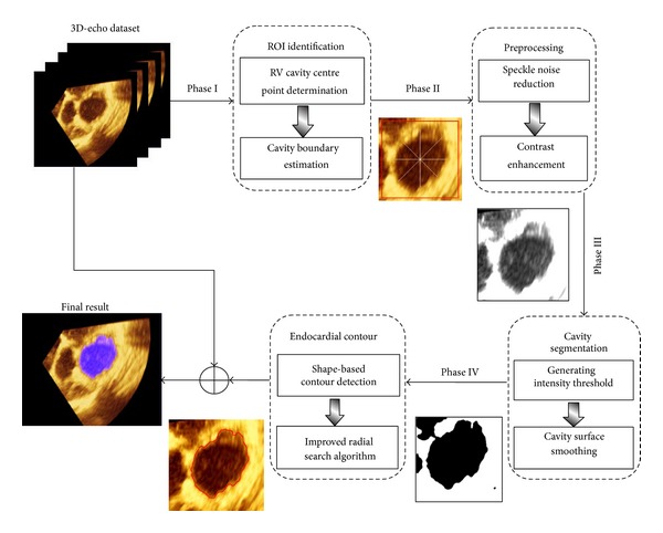
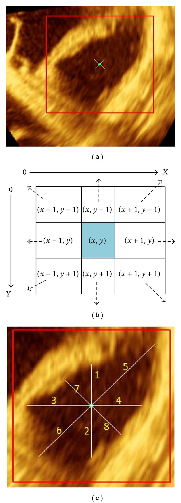
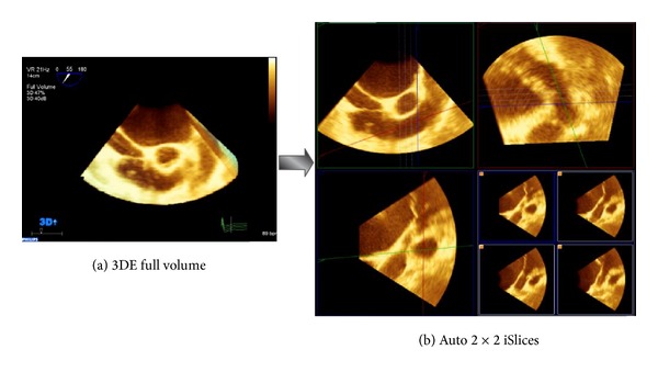
This paper presents a hybrid method to extract endocardial contour of the right ventricular (RV) in 4-slices from 3D echocardiography dataset. The overall framework comprises four processing phases. In Phase I, the region of interest (ROI) is identified by estimating the cavity boundary. Speckle noise reduction and contrast enhancement were implemented in Phase II as preprocessing tasks. In Phase III, the RV cavity region was segmented by generating intensity threshold which was used for once for all frames. Finally, Phase IV is proposed to extract the RV endocardial contour in a complete cardiac cycle using a combination of shape-based contour detection and improved radial search algorithm. The proposed method was applied to 16 datasets of 3D echocardiography encompassing the RV in long-axis view. The accuracy of experimental results obtained by the proposed method was evaluated qualitatively and quantitatively. It has been done by comparing the segmentation results of RV cavity based on endocardial contour extraction with the ground truth. The comparative analysis results show that the proposed method performs efficiently in all datasets with overall performance of 95% and the root mean square distances (RMSD) measure in terms of mean ± SD was found to be 2.21 ± 0.35 mm for RV endocardial contours.

 求助内容:
求助内容: 应助结果提醒方式:


