Moug Al-Bakri, Ase Krogh Rasmussen, Carsten Thomsen, Peter Bjerre Toft
{"title":"Graves眼病的眼眶体积测量:与甲状腺功能障碍视神经病变有关的肌肉和脂肪累及。","authors":"Moug Al-Bakri, Ase Krogh Rasmussen, Carsten Thomsen, Peter Bjerre Toft","doi":"10.1155/2014/435276","DOIUrl":null,"url":null,"abstract":"<p><p>Purpose. We wanted to investigate the relative significance of fat and muscle enlargement in the development of dysthyroid optic neuropathy (DON) in Graves' orbitopathy (GO). Methods. Preoperative coronal CT scans of 13 patients with and without DON who subsequently underwent orbital decompression were retrospectively analyzed. Thirteen patients imaged for unilateral orbital fractures served as controls. Results. The retrobulbar muscle volume was 2.1 ± 0.5 cm(3) (mean ± SD) in controls, 4.3 ± 1.5 cm(3) in GO without DON, and 4.7 ± 1.7 cm(3) in GO with DON. The retrobulbar fat volume was 5.4 ± 1.6 cm(3) in controls, 8.7 ± 8.0 cm(3) in GO without DON, and 9.4 ± 3.1 cm(3) in GO with DON. The muscle and fat volumes were higher in patients with GO than in controls (P < 0.001), but the volumes in orbits with and without DON were not significantly different. The volume of the optic nerve were similar in the 3 groups. The number of apical, coronal 2 mm thick slices with no fat was 2.9 ± 0.9 in normal orbits, it was 4.1 ± 1.0 in GO orbits without DON and 5.3 ± 0.8 in GO orbits with DON (P = 0.007). Conclusion. Apical muscle enlargement may be more important than orbital fat enlargement in the development of DON. However, the fact that apical crowding and muscle enlargement also occur in orbits without DON suggests that other factors also play a role in the development of DON. </p>","PeriodicalId":90193,"journal":{"name":"ISRN ophthalmology","volume":"2014 ","pages":"435276"},"PeriodicalIF":0.0000,"publicationDate":"2014-04-02","publicationTypes":"Journal Article","fieldsOfStudy":null,"isOpenAccess":false,"openAccessPdf":"https://sci-hub-pdf.com/10.1155/2014/435276","citationCount":"19","resultStr":"{\"title\":\"Orbital Volumetry in Graves' Orbitopathy: Muscle and Fat Involvement in relation to Dysthyroid Optic Neuropathy.\",\"authors\":\"Moug Al-Bakri, Ase Krogh Rasmussen, Carsten Thomsen, Peter Bjerre Toft\",\"doi\":\"10.1155/2014/435276\",\"DOIUrl\":null,\"url\":null,\"abstract\":\"<p><p>Purpose. We wanted to investigate the relative significance of fat and muscle enlargement in the development of dysthyroid optic neuropathy (DON) in Graves' orbitopathy (GO). Methods. Preoperative coronal CT scans of 13 patients with and without DON who subsequently underwent orbital decompression were retrospectively analyzed. Thirteen patients imaged for unilateral orbital fractures served as controls. Results. The retrobulbar muscle volume was 2.1 ± 0.5 cm(3) (mean ± SD) in controls, 4.3 ± 1.5 cm(3) in GO without DON, and 4.7 ± 1.7 cm(3) in GO with DON. The retrobulbar fat volume was 5.4 ± 1.6 cm(3) in controls, 8.7 ± 8.0 cm(3) in GO without DON, and 9.4 ± 3.1 cm(3) in GO with DON. The muscle and fat volumes were higher in patients with GO than in controls (P < 0.001), but the volumes in orbits with and without DON were not significantly different. The volume of the optic nerve were similar in the 3 groups. The number of apical, coronal 2 mm thick slices with no fat was 2.9 ± 0.9 in normal orbits, it was 4.1 ± 1.0 in GO orbits without DON and 5.3 ± 0.8 in GO orbits with DON (P = 0.007). Conclusion. Apical muscle enlargement may be more important than orbital fat enlargement in the development of DON. However, the fact that apical crowding and muscle enlargement also occur in orbits without DON suggests that other factors also play a role in the development of DON. </p>\",\"PeriodicalId\":90193,\"journal\":{\"name\":\"ISRN ophthalmology\",\"volume\":\"2014 \",\"pages\":\"435276\"},\"PeriodicalIF\":0.0000,\"publicationDate\":\"2014-04-02\",\"publicationTypes\":\"Journal Article\",\"fieldsOfStudy\":null,\"isOpenAccess\":false,\"openAccessPdf\":\"https://sci-hub-pdf.com/10.1155/2014/435276\",\"citationCount\":\"19\",\"resultStr\":null,\"platform\":\"Semanticscholar\",\"paperid\":null,\"PeriodicalName\":\"ISRN ophthalmology\",\"FirstCategoryId\":\"1085\",\"ListUrlMain\":\"https://doi.org/10.1155/2014/435276\",\"RegionNum\":0,\"RegionCategory\":null,\"ArticlePicture\":[],\"TitleCN\":null,\"AbstractTextCN\":null,\"PMCID\":null,\"EPubDate\":\"2014/1/1 0:00:00\",\"PubModel\":\"eCollection\",\"JCR\":\"\",\"JCRName\":\"\",\"Score\":null,\"Total\":0}","platform":"Semanticscholar","paperid":null,"PeriodicalName":"ISRN ophthalmology","FirstCategoryId":"1085","ListUrlMain":"https://doi.org/10.1155/2014/435276","RegionNum":0,"RegionCategory":null,"ArticlePicture":[],"TitleCN":null,"AbstractTextCN":null,"PMCID":null,"EPubDate":"2014/1/1 0:00:00","PubModel":"eCollection","JCR":"","JCRName":"","Score":null,"Total":0}
Orbital Volumetry in Graves' Orbitopathy: Muscle and Fat Involvement in relation to Dysthyroid Optic Neuropathy.
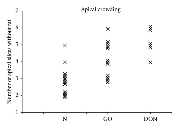
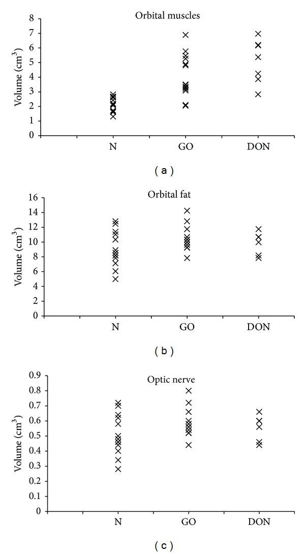
Purpose. We wanted to investigate the relative significance of fat and muscle enlargement in the development of dysthyroid optic neuropathy (DON) in Graves' orbitopathy (GO). Methods. Preoperative coronal CT scans of 13 patients with and without DON who subsequently underwent orbital decompression were retrospectively analyzed. Thirteen patients imaged for unilateral orbital fractures served as controls. Results. The retrobulbar muscle volume was 2.1 ± 0.5 cm(3) (mean ± SD) in controls, 4.3 ± 1.5 cm(3) in GO without DON, and 4.7 ± 1.7 cm(3) in GO with DON. The retrobulbar fat volume was 5.4 ± 1.6 cm(3) in controls, 8.7 ± 8.0 cm(3) in GO without DON, and 9.4 ± 3.1 cm(3) in GO with DON. The muscle and fat volumes were higher in patients with GO than in controls (P < 0.001), but the volumes in orbits with and without DON were not significantly different. The volume of the optic nerve were similar in the 3 groups. The number of apical, coronal 2 mm thick slices with no fat was 2.9 ± 0.9 in normal orbits, it was 4.1 ± 1.0 in GO orbits without DON and 5.3 ± 0.8 in GO orbits with DON (P = 0.007). Conclusion. Apical muscle enlargement may be more important than orbital fat enlargement in the development of DON. However, the fact that apical crowding and muscle enlargement also occur in orbits without DON suggests that other factors also play a role in the development of DON.

 求助内容:
求助内容: 应助结果提醒方式:


