Seung-Hyun Hong, Xi Jiang, Li Chen, Pujan Josh, Dong-Guk Shin, David Rowe
{"title":"计算机自动静态,动态和细胞骨组织形态测定。","authors":"Seung-Hyun Hong, Xi Jiang, Li Chen, Pujan Josh, Dong-Guk Shin, David Rowe","doi":"10.4172/2157-7552.S1-004","DOIUrl":null,"url":null,"abstract":"<p><p>Dynamic and cellular histomorphometry of trabeculae is the most biologically relevant way of assessing steady state bone health. Traditional measurement involves manual visual feature identification by a trained and qualified professional. Inherent with this methodology is the time and cost expenditure, as well as the subjectivity that naturally arises under human visual inspection. In this work, we propose a rapidly deployable, automated, and objective method for dynamic histomorphometry. We demonstrate that our method is highly effective in assessing cellular activities in distal femur and vertebra of mice which are injected with calcein and alizarin complexone 7 and 2 days prior to sacrifice. The mineralized bone tissues of mice are cryosectioned using a tape transfer protocol. A sequential workflow is implemented in which endogenous fluorescent signals (bone mineral, green and red mineralization lines), tartrate resistant acid phosphatase identified by ELF-97 and alkaline phosphatase identified by Fast Red are captured as individual tiled images of the section for each fluorescent color. All the images are then submitted to an image analysis pipeline that automates identification of the mineralized regions of bone and selection of a region of interest. The TRAP and AP stained images are aligned to the mineralized image using strategically placed fluorescent registration beads. Fluorescent signals are identified and are related to the trabecular surface within the ROI. Subsequently, the pipelined method computes static measurements, dynamic measurements, and cellular activities of osteoclast and osteoblast related to the trabecular surface. Our method has been applied to the distal femurs and vertebrae of 8 and 16 week old male and female C57Bl/6J mice. The histomorphometric results reveal a significantly greater bone turnover rate in female in contrast to male irrespective of age, validating similar outcomes reported by other studies.</p>","PeriodicalId":89595,"journal":{"name":"Journal of tissue science & engineering","volume":"Suppl 1 ","pages":"004"},"PeriodicalIF":0.0000,"publicationDate":"2012-12-24","publicationTypes":"Journal Article","fieldsOfStudy":null,"isOpenAccess":false,"openAccessPdf":"https://www.ncbi.nlm.nih.gov/pmc/articles/PMC4090931/pdf/","citationCount":"30","resultStr":"{\"title\":\"Computer-Automated Static, Dynamic and Cellular Bone Histomorphometry.\",\"authors\":\"Seung-Hyun Hong, Xi Jiang, Li Chen, Pujan Josh, Dong-Guk Shin, David Rowe\",\"doi\":\"10.4172/2157-7552.S1-004\",\"DOIUrl\":null,\"url\":null,\"abstract\":\"<p><p>Dynamic and cellular histomorphometry of trabeculae is the most biologically relevant way of assessing steady state bone health. Traditional measurement involves manual visual feature identification by a trained and qualified professional. Inherent with this methodology is the time and cost expenditure, as well as the subjectivity that naturally arises under human visual inspection. In this work, we propose a rapidly deployable, automated, and objective method for dynamic histomorphometry. We demonstrate that our method is highly effective in assessing cellular activities in distal femur and vertebra of mice which are injected with calcein and alizarin complexone 7 and 2 days prior to sacrifice. The mineralized bone tissues of mice are cryosectioned using a tape transfer protocol. A sequential workflow is implemented in which endogenous fluorescent signals (bone mineral, green and red mineralization lines), tartrate resistant acid phosphatase identified by ELF-97 and alkaline phosphatase identified by Fast Red are captured as individual tiled images of the section for each fluorescent color. All the images are then submitted to an image analysis pipeline that automates identification of the mineralized regions of bone and selection of a region of interest. The TRAP and AP stained images are aligned to the mineralized image using strategically placed fluorescent registration beads. Fluorescent signals are identified and are related to the trabecular surface within the ROI. Subsequently, the pipelined method computes static measurements, dynamic measurements, and cellular activities of osteoclast and osteoblast related to the trabecular surface. Our method has been applied to the distal femurs and vertebrae of 8 and 16 week old male and female C57Bl/6J mice. The histomorphometric results reveal a significantly greater bone turnover rate in female in contrast to male irrespective of age, validating similar outcomes reported by other studies.</p>\",\"PeriodicalId\":89595,\"journal\":{\"name\":\"Journal of tissue science & engineering\",\"volume\":\"Suppl 1 \",\"pages\":\"004\"},\"PeriodicalIF\":0.0000,\"publicationDate\":\"2012-12-24\",\"publicationTypes\":\"Journal Article\",\"fieldsOfStudy\":null,\"isOpenAccess\":false,\"openAccessPdf\":\"https://www.ncbi.nlm.nih.gov/pmc/articles/PMC4090931/pdf/\",\"citationCount\":\"30\",\"resultStr\":null,\"platform\":\"Semanticscholar\",\"paperid\":null,\"PeriodicalName\":\"Journal of tissue science & engineering\",\"FirstCategoryId\":\"1085\",\"ListUrlMain\":\"https://doi.org/10.4172/2157-7552.S1-004\",\"RegionNum\":0,\"RegionCategory\":null,\"ArticlePicture\":[],\"TitleCN\":null,\"AbstractTextCN\":null,\"PMCID\":null,\"EPubDate\":\"\",\"PubModel\":\"\",\"JCR\":\"\",\"JCRName\":\"\",\"Score\":null,\"Total\":0}","platform":"Semanticscholar","paperid":null,"PeriodicalName":"Journal of tissue science & engineering","FirstCategoryId":"1085","ListUrlMain":"https://doi.org/10.4172/2157-7552.S1-004","RegionNum":0,"RegionCategory":null,"ArticlePicture":[],"TitleCN":null,"AbstractTextCN":null,"PMCID":null,"EPubDate":"","PubModel":"","JCR":"","JCRName":"","Score":null,"Total":0}
Computer-Automated Static, Dynamic and Cellular Bone Histomorphometry.
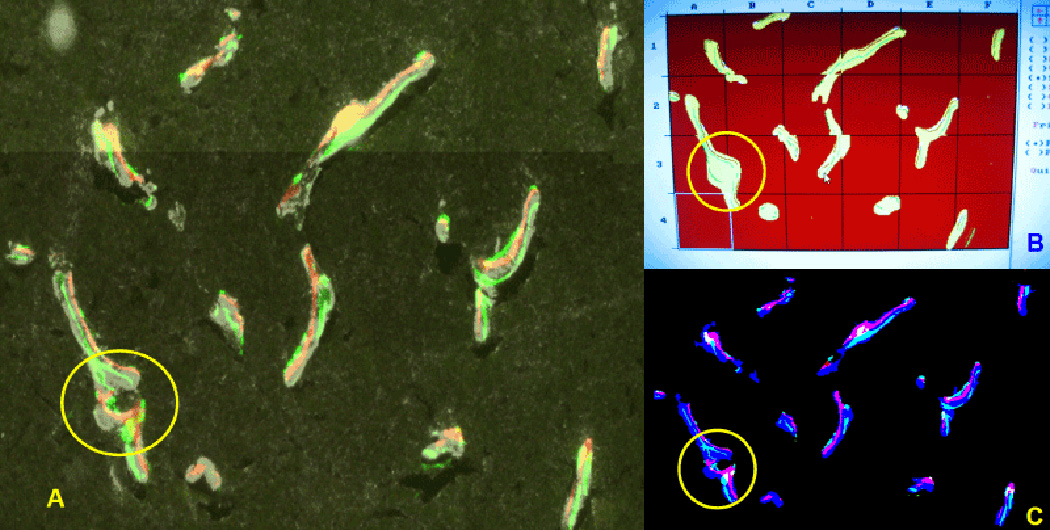
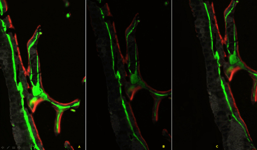
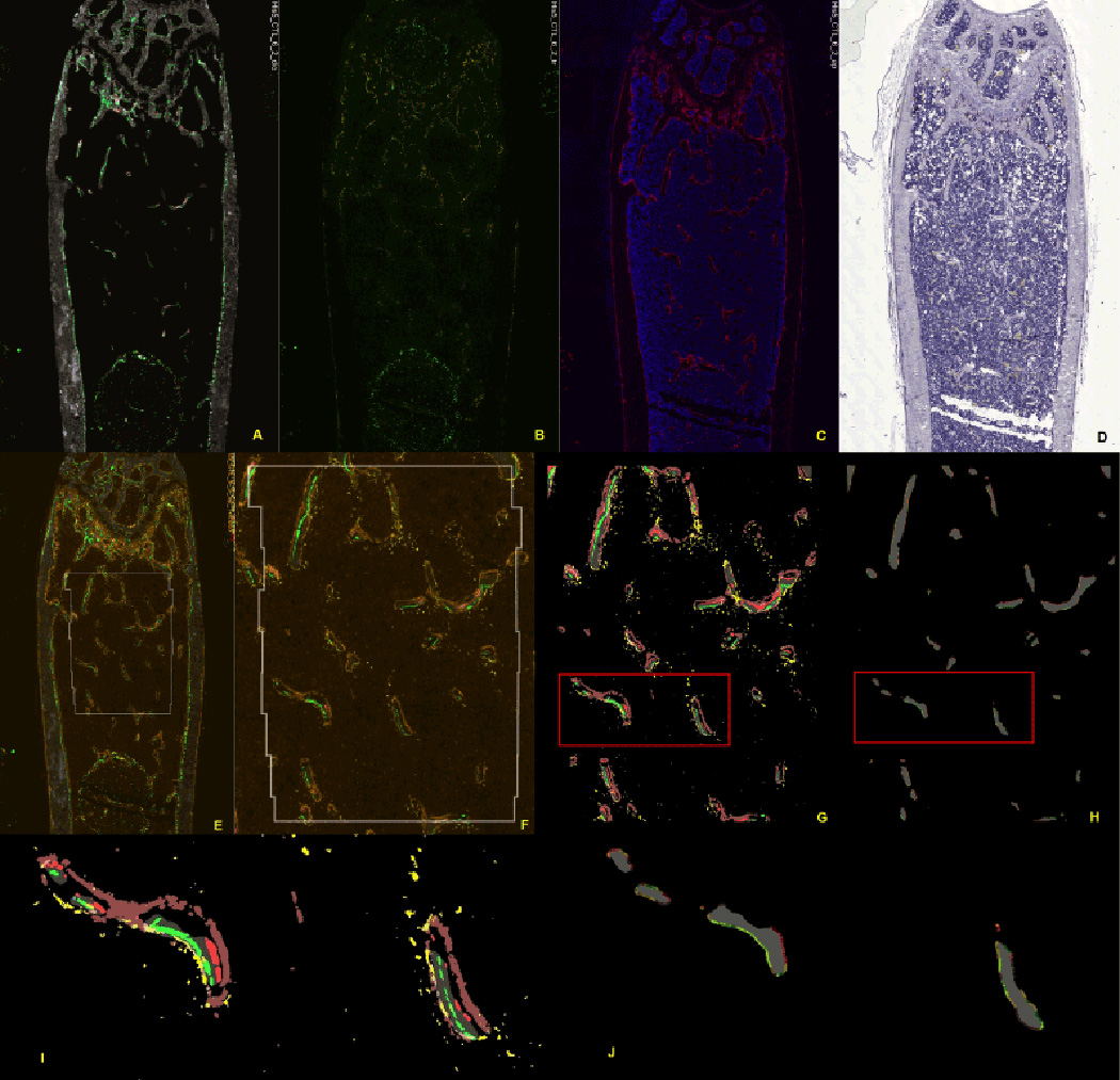
Dynamic and cellular histomorphometry of trabeculae is the most biologically relevant way of assessing steady state bone health. Traditional measurement involves manual visual feature identification by a trained and qualified professional. Inherent with this methodology is the time and cost expenditure, as well as the subjectivity that naturally arises under human visual inspection. In this work, we propose a rapidly deployable, automated, and objective method for dynamic histomorphometry. We demonstrate that our method is highly effective in assessing cellular activities in distal femur and vertebra of mice which are injected with calcein and alizarin complexone 7 and 2 days prior to sacrifice. The mineralized bone tissues of mice are cryosectioned using a tape transfer protocol. A sequential workflow is implemented in which endogenous fluorescent signals (bone mineral, green and red mineralization lines), tartrate resistant acid phosphatase identified by ELF-97 and alkaline phosphatase identified by Fast Red are captured as individual tiled images of the section for each fluorescent color. All the images are then submitted to an image analysis pipeline that automates identification of the mineralized regions of bone and selection of a region of interest. The TRAP and AP stained images are aligned to the mineralized image using strategically placed fluorescent registration beads. Fluorescent signals are identified and are related to the trabecular surface within the ROI. Subsequently, the pipelined method computes static measurements, dynamic measurements, and cellular activities of osteoclast and osteoblast related to the trabecular surface. Our method has been applied to the distal femurs and vertebrae of 8 and 16 week old male and female C57Bl/6J mice. The histomorphometric results reveal a significantly greater bone turnover rate in female in contrast to male irrespective of age, validating similar outcomes reported by other studies.

 求助内容:
求助内容: 应助结果提醒方式:


