{"title":"银结合的核仁组织区在伴有上皮发育不良和口腔鳞状细胞癌的口腔白斑中出现斑点:一项体内研究。","authors":"Fahad Mansoor Samadi, Bastain Thattil Sebastian, Anil Singh, Shaleen Chandra, Shadab Mohammad, Arun Singh, Thippeswamy Halappa, Firoza Samadi","doi":"10.1155/2014/479187","DOIUrl":null,"url":null,"abstract":"<p><p>Silver binding nucleolar organizer regions (AgNOR) in normal oral mucosa (NOM), oral leukoplakia with epithelial dysplasia (ED), and oral squamous cell carcinoma (OSCC) were studied. The mean AgNOR count per nucleus increased from NOM to ED to OSCC. Tissue showing ED in oral leukoplakia and OSCC cases showed higher counts, wider scatter, and smaller size of AgNOR dots in the nuclei. The study seems to suggest that time method has some potential in distinguishing between NOM and oral leukoplakia with ED and OSCC. Studies of larger numbers are needed to arrive at more substantial conclusions. </p>","PeriodicalId":89396,"journal":{"name":"ISRN dentistry","volume":"2014 ","pages":"479187"},"PeriodicalIF":0.0000,"publicationDate":"2014-04-30","publicationTypes":"Journal Article","fieldsOfStudy":null,"isOpenAccess":false,"openAccessPdf":"https://sci-hub-pdf.com/10.1155/2014/479187","citationCount":"9","resultStr":"{\"title\":\"Silver binding nucleolar organizer regions dots in oral leukoplakia with epithelial dysplasia and oral squamous cell carcinoma: an in vivo study.\",\"authors\":\"Fahad Mansoor Samadi, Bastain Thattil Sebastian, Anil Singh, Shaleen Chandra, Shadab Mohammad, Arun Singh, Thippeswamy Halappa, Firoza Samadi\",\"doi\":\"10.1155/2014/479187\",\"DOIUrl\":null,\"url\":null,\"abstract\":\"<p><p>Silver binding nucleolar organizer regions (AgNOR) in normal oral mucosa (NOM), oral leukoplakia with epithelial dysplasia (ED), and oral squamous cell carcinoma (OSCC) were studied. The mean AgNOR count per nucleus increased from NOM to ED to OSCC. Tissue showing ED in oral leukoplakia and OSCC cases showed higher counts, wider scatter, and smaller size of AgNOR dots in the nuclei. The study seems to suggest that time method has some potential in distinguishing between NOM and oral leukoplakia with ED and OSCC. Studies of larger numbers are needed to arrive at more substantial conclusions. </p>\",\"PeriodicalId\":89396,\"journal\":{\"name\":\"ISRN dentistry\",\"volume\":\"2014 \",\"pages\":\"479187\"},\"PeriodicalIF\":0.0000,\"publicationDate\":\"2014-04-30\",\"publicationTypes\":\"Journal Article\",\"fieldsOfStudy\":null,\"isOpenAccess\":false,\"openAccessPdf\":\"https://sci-hub-pdf.com/10.1155/2014/479187\",\"citationCount\":\"9\",\"resultStr\":null,\"platform\":\"Semanticscholar\",\"paperid\":null,\"PeriodicalName\":\"ISRN dentistry\",\"FirstCategoryId\":\"1085\",\"ListUrlMain\":\"https://doi.org/10.1155/2014/479187\",\"RegionNum\":0,\"RegionCategory\":null,\"ArticlePicture\":[],\"TitleCN\":null,\"AbstractTextCN\":null,\"PMCID\":null,\"EPubDate\":\"2014/1/1 0:00:00\",\"PubModel\":\"eCollection\",\"JCR\":\"\",\"JCRName\":\"\",\"Score\":null,\"Total\":0}","platform":"Semanticscholar","paperid":null,"PeriodicalName":"ISRN dentistry","FirstCategoryId":"1085","ListUrlMain":"https://doi.org/10.1155/2014/479187","RegionNum":0,"RegionCategory":null,"ArticlePicture":[],"TitleCN":null,"AbstractTextCN":null,"PMCID":null,"EPubDate":"2014/1/1 0:00:00","PubModel":"eCollection","JCR":"","JCRName":"","Score":null,"Total":0}
Silver binding nucleolar organizer regions dots in oral leukoplakia with epithelial dysplasia and oral squamous cell carcinoma: an in vivo study.
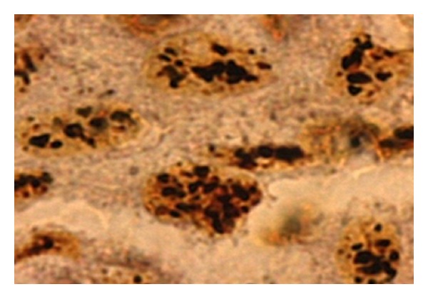
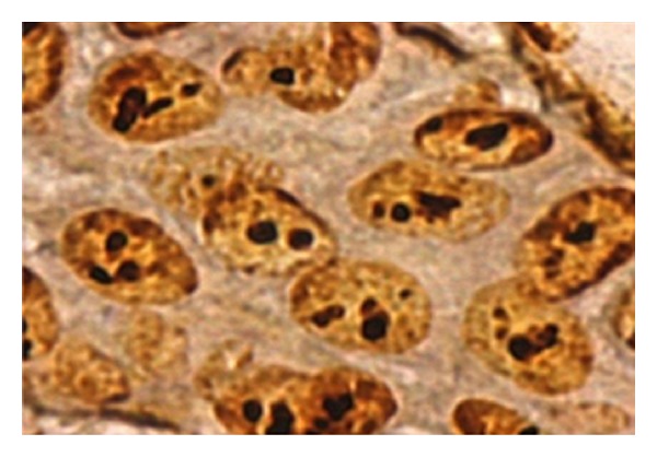
Silver binding nucleolar organizer regions (AgNOR) in normal oral mucosa (NOM), oral leukoplakia with epithelial dysplasia (ED), and oral squamous cell carcinoma (OSCC) were studied. The mean AgNOR count per nucleus increased from NOM to ED to OSCC. Tissue showing ED in oral leukoplakia and OSCC cases showed higher counts, wider scatter, and smaller size of AgNOR dots in the nuclei. The study seems to suggest that time method has some potential in distinguishing between NOM and oral leukoplakia with ED and OSCC. Studies of larger numbers are needed to arrive at more substantial conclusions.

 求助内容:
求助内容: 应助结果提醒方式:


