Stephanie Boehme, Alexander Mohr, Michael Pi Becker, Wolfgang Hr Miltner, Thomas Straube
{"title":"社交焦虑障碍视频诱发症状时脑激活的区域依赖时间过程。","authors":"Stephanie Boehme, Alexander Mohr, Michael Pi Becker, Wolfgang Hr Miltner, Thomas Straube","doi":"10.1186/2045-5380-4-6","DOIUrl":null,"url":null,"abstract":"<p><strong>Background: </strong>Previous functional imaging studies using symptom provocation in patients with social anxiety disorder (SAD) reported inconsistent findings, which might be at least partially related to different time-dependent activation profiles in different brain areas. In the present functional magnetic resonance imaging study, we used a novel video-based symptom provocation design in order to investigate the magnitude and time course of activation in different brain areas in 20 SAD patients and 20 healthy controls.</p><p><strong>Results: </strong>The disorder-related videos induced increased anxiety in patients with SAD as compared to healthy controls. Analyses of brain activation to disorder-related versus neutral video clips revealed amygdala activation during the first but not during the second half of the clips in patients as compared to controls. In contrast, the activation in the insula showed a reversed pattern with increased activation during the second but not during the first half of the video clips. Furthermore, a cluster in the anterior dorsal anterior cingulate cortex showed a sustained response for the entire duration of the videos.</p><p><strong>Conclusions: </strong>The present findings suggest that different regions of the fear network show differential temporal response patterns during video-induced symptom provocation in SAD. While the amygdala is involved during initial threat processing, the insula seems to be more involved during subsequent anxiety responses. In accordance with cognitive models of SAD, a medial prefrontal region engaged in emotional-cognitive interactions is generally hyperactivated.</p>","PeriodicalId":89532,"journal":{"name":"Biology of mood & anxiety disorders","volume":"4 ","pages":"6"},"PeriodicalIF":0.0000,"publicationDate":"2014-04-28","publicationTypes":"Journal Article","fieldsOfStudy":null,"isOpenAccess":false,"openAccessPdf":"https://sci-hub-pdf.com/10.1186/2045-5380-4-6","citationCount":"21","resultStr":"{\"title\":\"Area-dependent time courses of brain activation during video-induced symptom provocation in social anxiety disorder.\",\"authors\":\"Stephanie Boehme, Alexander Mohr, Michael Pi Becker, Wolfgang Hr Miltner, Thomas Straube\",\"doi\":\"10.1186/2045-5380-4-6\",\"DOIUrl\":null,\"url\":null,\"abstract\":\"<p><strong>Background: </strong>Previous functional imaging studies using symptom provocation in patients with social anxiety disorder (SAD) reported inconsistent findings, which might be at least partially related to different time-dependent activation profiles in different brain areas. In the present functional magnetic resonance imaging study, we used a novel video-based symptom provocation design in order to investigate the magnitude and time course of activation in different brain areas in 20 SAD patients and 20 healthy controls.</p><p><strong>Results: </strong>The disorder-related videos induced increased anxiety in patients with SAD as compared to healthy controls. Analyses of brain activation to disorder-related versus neutral video clips revealed amygdala activation during the first but not during the second half of the clips in patients as compared to controls. In contrast, the activation in the insula showed a reversed pattern with increased activation during the second but not during the first half of the video clips. Furthermore, a cluster in the anterior dorsal anterior cingulate cortex showed a sustained response for the entire duration of the videos.</p><p><strong>Conclusions: </strong>The present findings suggest that different regions of the fear network show differential temporal response patterns during video-induced symptom provocation in SAD. While the amygdala is involved during initial threat processing, the insula seems to be more involved during subsequent anxiety responses. In accordance with cognitive models of SAD, a medial prefrontal region engaged in emotional-cognitive interactions is generally hyperactivated.</p>\",\"PeriodicalId\":89532,\"journal\":{\"name\":\"Biology of mood & anxiety disorders\",\"volume\":\"4 \",\"pages\":\"6\"},\"PeriodicalIF\":0.0000,\"publicationDate\":\"2014-04-28\",\"publicationTypes\":\"Journal Article\",\"fieldsOfStudy\":null,\"isOpenAccess\":false,\"openAccessPdf\":\"https://sci-hub-pdf.com/10.1186/2045-5380-4-6\",\"citationCount\":\"21\",\"resultStr\":null,\"platform\":\"Semanticscholar\",\"paperid\":null,\"PeriodicalName\":\"Biology of mood & anxiety disorders\",\"FirstCategoryId\":\"1085\",\"ListUrlMain\":\"https://doi.org/10.1186/2045-5380-4-6\",\"RegionNum\":0,\"RegionCategory\":null,\"ArticlePicture\":[],\"TitleCN\":null,\"AbstractTextCN\":null,\"PMCID\":null,\"EPubDate\":\"2014/1/1 0:00:00\",\"PubModel\":\"eCollection\",\"JCR\":\"\",\"JCRName\":\"\",\"Score\":null,\"Total\":0}","platform":"Semanticscholar","paperid":null,"PeriodicalName":"Biology of mood & anxiety disorders","FirstCategoryId":"1085","ListUrlMain":"https://doi.org/10.1186/2045-5380-4-6","RegionNum":0,"RegionCategory":null,"ArticlePicture":[],"TitleCN":null,"AbstractTextCN":null,"PMCID":null,"EPubDate":"2014/1/1 0:00:00","PubModel":"eCollection","JCR":"","JCRName":"","Score":null,"Total":0}
Area-dependent time courses of brain activation during video-induced symptom provocation in social anxiety disorder.
Background: Previous functional imaging studies using symptom provocation in patients with social anxiety disorder (SAD) reported inconsistent findings, which might be at least partially related to different time-dependent activation profiles in different brain areas. In the present functional magnetic resonance imaging study, we used a novel video-based symptom provocation design in order to investigate the magnitude and time course of activation in different brain areas in 20 SAD patients and 20 healthy controls.
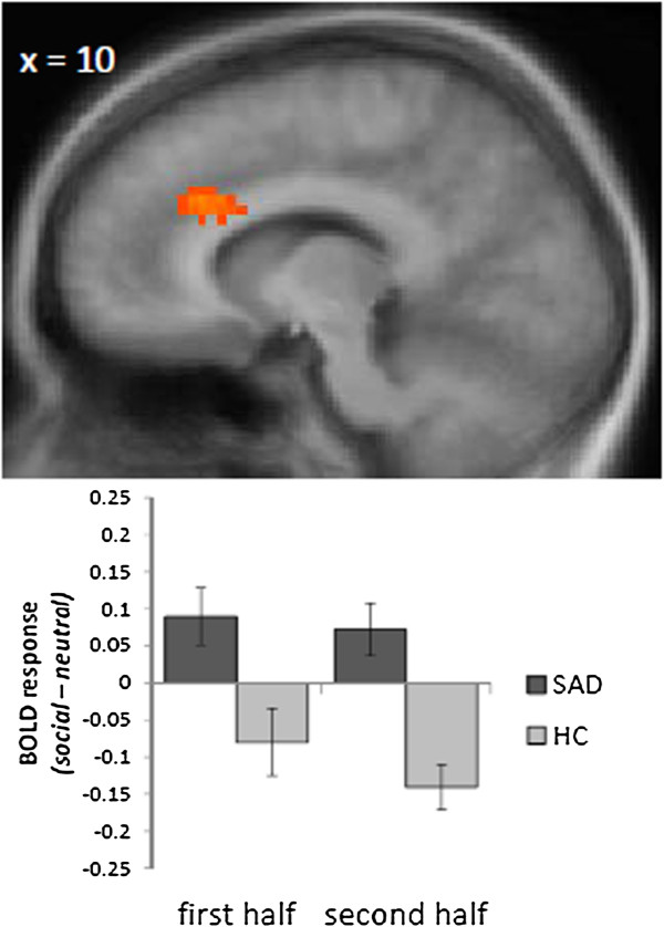
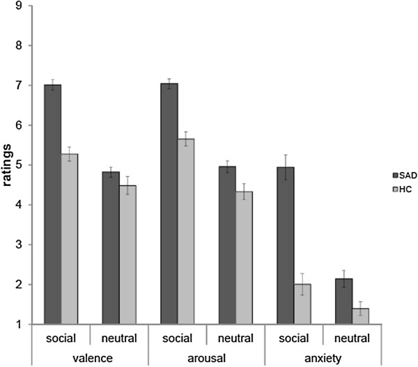
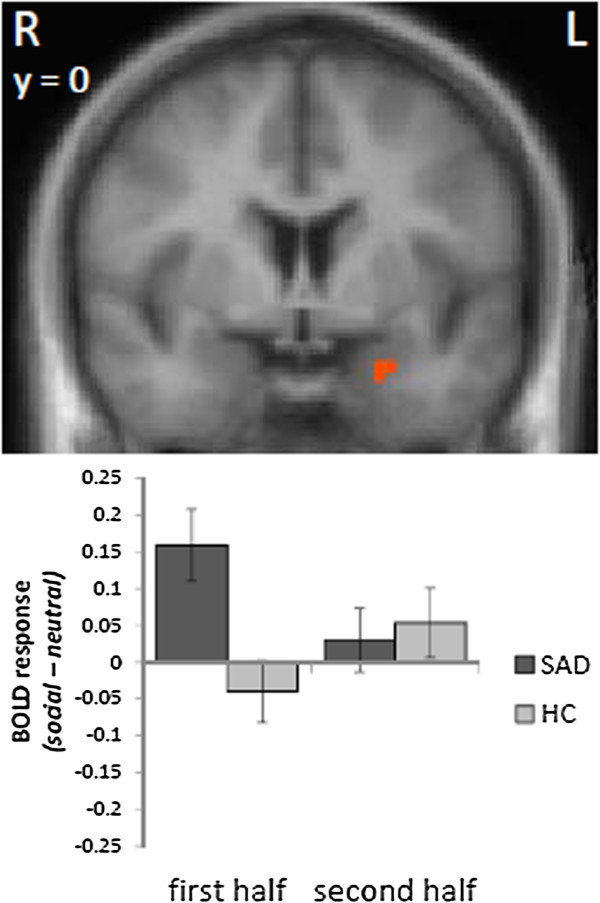
Results: The disorder-related videos induced increased anxiety in patients with SAD as compared to healthy controls. Analyses of brain activation to disorder-related versus neutral video clips revealed amygdala activation during the first but not during the second half of the clips in patients as compared to controls. In contrast, the activation in the insula showed a reversed pattern with increased activation during the second but not during the first half of the video clips. Furthermore, a cluster in the anterior dorsal anterior cingulate cortex showed a sustained response for the entire duration of the videos.
Conclusions: The present findings suggest that different regions of the fear network show differential temporal response patterns during video-induced symptom provocation in SAD. While the amygdala is involved during initial threat processing, the insula seems to be more involved during subsequent anxiety responses. In accordance with cognitive models of SAD, a medial prefrontal region engaged in emotional-cognitive interactions is generally hyperactivated.

 求助内容:
求助内容: 应助结果提醒方式:


