{"title":"软组织肿瘤组织细胞病理学相关性的前瞻性研究。","authors":"Priyanka Bhatia Soni, Anand Kumar Verma, Raj Kumar Chandoke, Jitendra Singh Nigam","doi":"10.1155/2014/678628","DOIUrl":null,"url":null,"abstract":"<p><p>Background. Soft tissue tumors are defined as nonepithelial extraskeletal tissue of the body exclusive of the reticuloendothelial system, glia, and supporting tissue of various parenchymal organs. The absence of recognizable tissue architectural patterns in cytological preparation makes diagnosis by FNAC more difficult. Aims. To assess the utility of FNAC in diagnosing soft tissue tumors and to determine their patterns compared with with the respective histopathology results. Materials and Methods. 150 cases of soft tissue tumors were included in this study for cytologic and histologic correlation. FNAC air dried smears were stained with Giemsa stain and 95% ethanol fixed smears were stained with Papanicolaou stain. The smears were studied for cytological diagnosis and were categorized as benign, suspicious of malignancy, and malignant along with specific subtyping of the lesion. All diagnostic FNAC results were compared for diagnostic concordance using histology results as the \"gold standard.\" Results. The sensitivity, specificity, positive predictive value, negative predictive value, and efficiency were 70%, 100%, 97.90%, 100%, and 98%, respectively. P value was <0.0001 which shows statistically extreme significant correlation. Conclusion. FNAC is a very important preliminary diagnostic tool in palpable soft tissue lumps with high degree of correlation with the final histopathology report. </p>","PeriodicalId":89212,"journal":{"name":"Pathology research international","volume":"2014 ","pages":"678628"},"PeriodicalIF":0.0000,"publicationDate":"2014-01-01","publicationTypes":"Journal Article","fieldsOfStudy":null,"isOpenAccess":false,"openAccessPdf":"https://sci-hub-pdf.com/10.1155/2014/678628","citationCount":"22","resultStr":"{\"title\":\"A prospective study of soft tissue tumors histocytopathology correlation.\",\"authors\":\"Priyanka Bhatia Soni, Anand Kumar Verma, Raj Kumar Chandoke, Jitendra Singh Nigam\",\"doi\":\"10.1155/2014/678628\",\"DOIUrl\":null,\"url\":null,\"abstract\":\"<p><p>Background. Soft tissue tumors are defined as nonepithelial extraskeletal tissue of the body exclusive of the reticuloendothelial system, glia, and supporting tissue of various parenchymal organs. The absence of recognizable tissue architectural patterns in cytological preparation makes diagnosis by FNAC more difficult. Aims. To assess the utility of FNAC in diagnosing soft tissue tumors and to determine their patterns compared with with the respective histopathology results. Materials and Methods. 150 cases of soft tissue tumors were included in this study for cytologic and histologic correlation. FNAC air dried smears were stained with Giemsa stain and 95% ethanol fixed smears were stained with Papanicolaou stain. The smears were studied for cytological diagnosis and were categorized as benign, suspicious of malignancy, and malignant along with specific subtyping of the lesion. All diagnostic FNAC results were compared for diagnostic concordance using histology results as the \\\"gold standard.\\\" Results. The sensitivity, specificity, positive predictive value, negative predictive value, and efficiency were 70%, 100%, 97.90%, 100%, and 98%, respectively. P value was <0.0001 which shows statistically extreme significant correlation. Conclusion. FNAC is a very important preliminary diagnostic tool in palpable soft tissue lumps with high degree of correlation with the final histopathology report. </p>\",\"PeriodicalId\":89212,\"journal\":{\"name\":\"Pathology research international\",\"volume\":\"2014 \",\"pages\":\"678628\"},\"PeriodicalIF\":0.0000,\"publicationDate\":\"2014-01-01\",\"publicationTypes\":\"Journal Article\",\"fieldsOfStudy\":null,\"isOpenAccess\":false,\"openAccessPdf\":\"https://sci-hub-pdf.com/10.1155/2014/678628\",\"citationCount\":\"22\",\"resultStr\":null,\"platform\":\"Semanticscholar\",\"paperid\":null,\"PeriodicalName\":\"Pathology research international\",\"FirstCategoryId\":\"1085\",\"ListUrlMain\":\"https://doi.org/10.1155/2014/678628\",\"RegionNum\":0,\"RegionCategory\":null,\"ArticlePicture\":[],\"TitleCN\":null,\"AbstractTextCN\":null,\"PMCID\":null,\"EPubDate\":\"2014/4/28 0:00:00\",\"PubModel\":\"Epub\",\"JCR\":\"\",\"JCRName\":\"\",\"Score\":null,\"Total\":0}","platform":"Semanticscholar","paperid":null,"PeriodicalName":"Pathology research international","FirstCategoryId":"1085","ListUrlMain":"https://doi.org/10.1155/2014/678628","RegionNum":0,"RegionCategory":null,"ArticlePicture":[],"TitleCN":null,"AbstractTextCN":null,"PMCID":null,"EPubDate":"2014/4/28 0:00:00","PubModel":"Epub","JCR":"","JCRName":"","Score":null,"Total":0}
A prospective study of soft tissue tumors histocytopathology correlation.
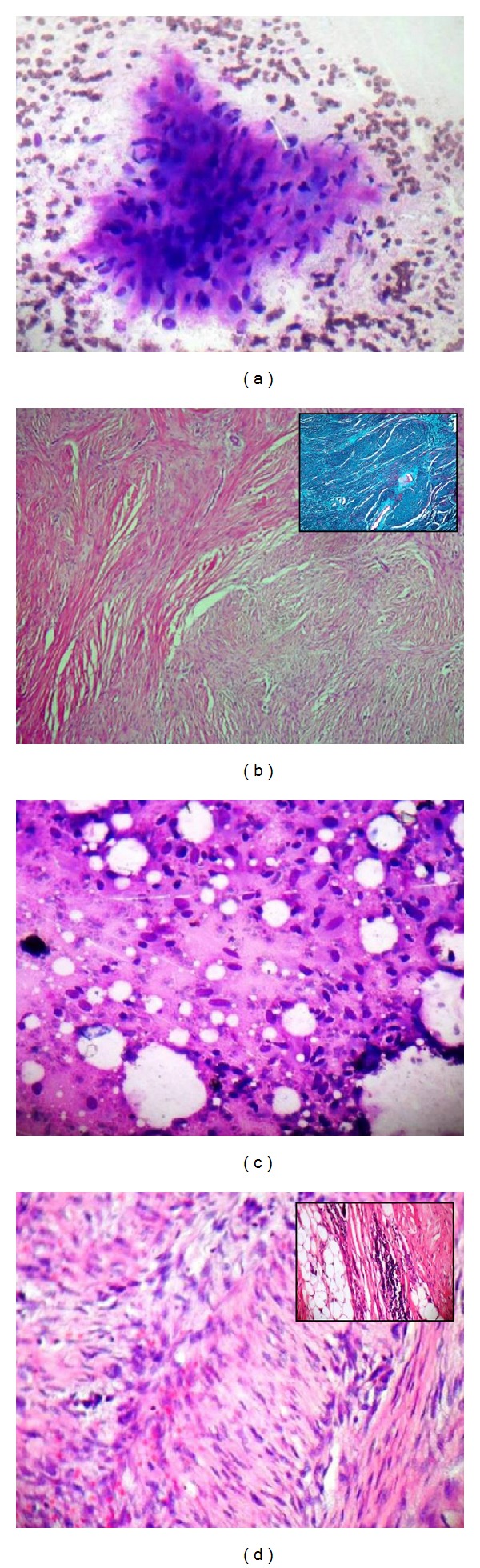
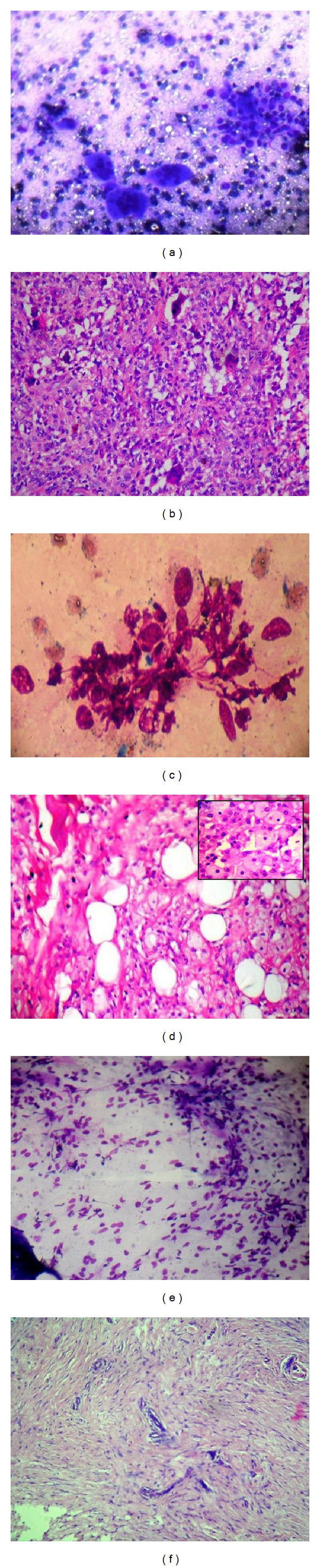
Background. Soft tissue tumors are defined as nonepithelial extraskeletal tissue of the body exclusive of the reticuloendothelial system, glia, and supporting tissue of various parenchymal organs. The absence of recognizable tissue architectural patterns in cytological preparation makes diagnosis by FNAC more difficult. Aims. To assess the utility of FNAC in diagnosing soft tissue tumors and to determine their patterns compared with with the respective histopathology results. Materials and Methods. 150 cases of soft tissue tumors were included in this study for cytologic and histologic correlation. FNAC air dried smears were stained with Giemsa stain and 95% ethanol fixed smears were stained with Papanicolaou stain. The smears were studied for cytological diagnosis and were categorized as benign, suspicious of malignancy, and malignant along with specific subtyping of the lesion. All diagnostic FNAC results were compared for diagnostic concordance using histology results as the "gold standard." Results. The sensitivity, specificity, positive predictive value, negative predictive value, and efficiency were 70%, 100%, 97.90%, 100%, and 98%, respectively. P value was <0.0001 which shows statistically extreme significant correlation. Conclusion. FNAC is a very important preliminary diagnostic tool in palpable soft tissue lumps with high degree of correlation with the final histopathology report.


 求助内容:
求助内容: 应助结果提醒方式:


