{"title":"伊拉克免疫正常小鼠和免疫抑制小鼠实验性视网膜脑虫感染的病理研究。","authors":"Hafidh I Al-Sadi, Saevan S Al-Mahmood","doi":"10.1155/2014/857036","DOIUrl":null,"url":null,"abstract":"<p><p>This study was performed to evaluate pathology of experimental Encephalitozoon cuniculi (Iraqi isolate) infection in normal and immunosuppressed mice. Pathological changes were not seen in negative control mice while secondary bacterial infections were noted in the lungs, kidneys, and heart of mice given dexamethasone. Typical E. cuniculi infection lesions were found in brain, livers, lungs, and kidneys of mice given 10(7) E. cuniculi spores/mouse orally. These lesions were in the form of nonsuppurative meningoencephalitis with vasculitis in brain, interstitial inflammation with infiltration of both lymphocytes and plasma cells in lung tissue, and nonsuppurative interstitial (focal and diffuse) nephritis, presence of vacuole containing mature and immature spores in enterocytes within the tips of villi, and lymphoiod hyperplasia of the white pulp and vasculitis of the intratrabecular vessels. Mice that were given 10(7) E. cuniculi spores/mouse orally showed lesions similar to those observed in the previous group (vasculitis and granulomas) but the lesions were more severe and widespread. In conclusion, this is the first report of experimental E. cuniculi infection induced by E. cuniculi isolated from a naturally infected rabbit in Iraq and that infection became more severe and widespread upon the administration of dexaethasone. </p>","PeriodicalId":89212,"journal":{"name":"Pathology research international","volume":"2014 ","pages":"857036"},"PeriodicalIF":0.0000,"publicationDate":"2014-01-01","publicationTypes":"Journal Article","fieldsOfStudy":null,"isOpenAccess":false,"openAccessPdf":"https://sci-hub-pdf.com/10.1155/2014/857036","citationCount":"14","resultStr":"{\"title\":\"Pathology of Experimental Encephalitozoon cuniculi Infection in Immunocompetent and Immunosuppressed Mice in Iraq.\",\"authors\":\"Hafidh I Al-Sadi, Saevan S Al-Mahmood\",\"doi\":\"10.1155/2014/857036\",\"DOIUrl\":null,\"url\":null,\"abstract\":\"<p><p>This study was performed to evaluate pathology of experimental Encephalitozoon cuniculi (Iraqi isolate) infection in normal and immunosuppressed mice. Pathological changes were not seen in negative control mice while secondary bacterial infections were noted in the lungs, kidneys, and heart of mice given dexamethasone. Typical E. cuniculi infection lesions were found in brain, livers, lungs, and kidneys of mice given 10(7) E. cuniculi spores/mouse orally. These lesions were in the form of nonsuppurative meningoencephalitis with vasculitis in brain, interstitial inflammation with infiltration of both lymphocytes and plasma cells in lung tissue, and nonsuppurative interstitial (focal and diffuse) nephritis, presence of vacuole containing mature and immature spores in enterocytes within the tips of villi, and lymphoiod hyperplasia of the white pulp and vasculitis of the intratrabecular vessels. Mice that were given 10(7) E. cuniculi spores/mouse orally showed lesions similar to those observed in the previous group (vasculitis and granulomas) but the lesions were more severe and widespread. In conclusion, this is the first report of experimental E. cuniculi infection induced by E. cuniculi isolated from a naturally infected rabbit in Iraq and that infection became more severe and widespread upon the administration of dexaethasone. </p>\",\"PeriodicalId\":89212,\"journal\":{\"name\":\"Pathology research international\",\"volume\":\"2014 \",\"pages\":\"857036\"},\"PeriodicalIF\":0.0000,\"publicationDate\":\"2014-01-01\",\"publicationTypes\":\"Journal Article\",\"fieldsOfStudy\":null,\"isOpenAccess\":false,\"openAccessPdf\":\"https://sci-hub-pdf.com/10.1155/2014/857036\",\"citationCount\":\"14\",\"resultStr\":null,\"platform\":\"Semanticscholar\",\"paperid\":null,\"PeriodicalName\":\"Pathology research international\",\"FirstCategoryId\":\"1085\",\"ListUrlMain\":\"https://doi.org/10.1155/2014/857036\",\"RegionNum\":0,\"RegionCategory\":null,\"ArticlePicture\":[],\"TitleCN\":null,\"AbstractTextCN\":null,\"PMCID\":null,\"EPubDate\":\"2014/3/20 0:00:00\",\"PubModel\":\"Epub\",\"JCR\":\"\",\"JCRName\":\"\",\"Score\":null,\"Total\":0}","platform":"Semanticscholar","paperid":null,"PeriodicalName":"Pathology research international","FirstCategoryId":"1085","ListUrlMain":"https://doi.org/10.1155/2014/857036","RegionNum":0,"RegionCategory":null,"ArticlePicture":[],"TitleCN":null,"AbstractTextCN":null,"PMCID":null,"EPubDate":"2014/3/20 0:00:00","PubModel":"Epub","JCR":"","JCRName":"","Score":null,"Total":0}
Pathology of Experimental Encephalitozoon cuniculi Infection in Immunocompetent and Immunosuppressed Mice in Iraq.
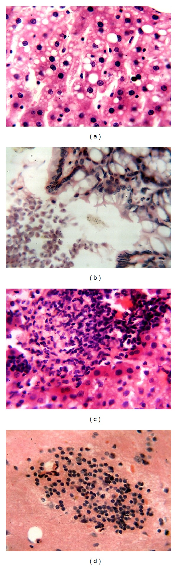
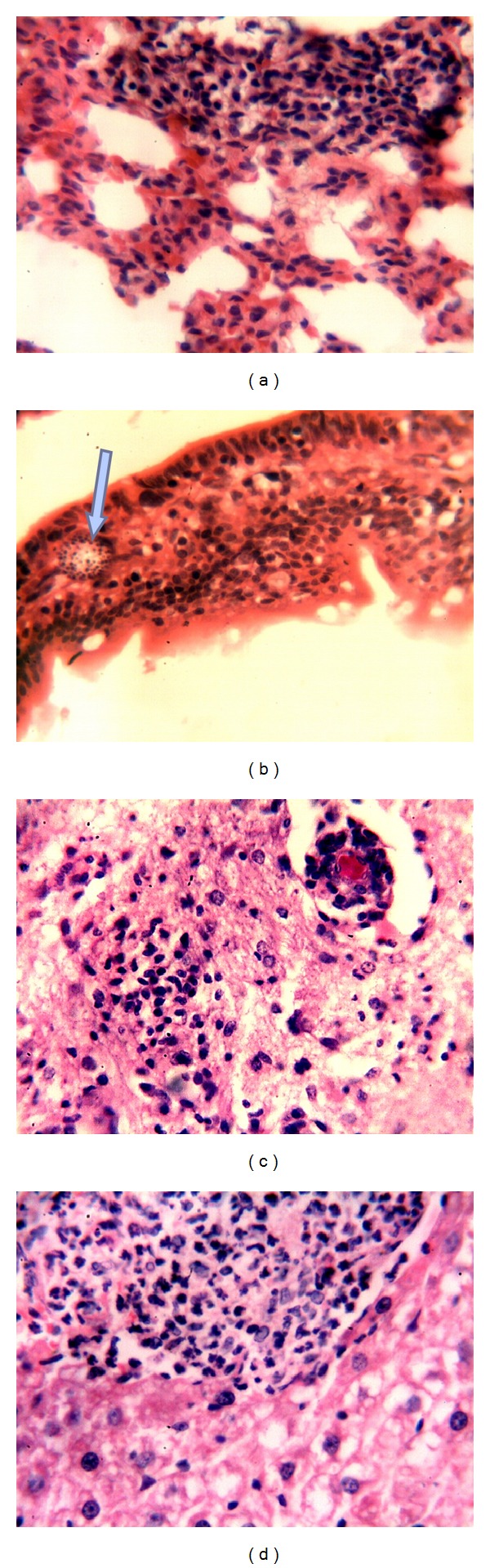
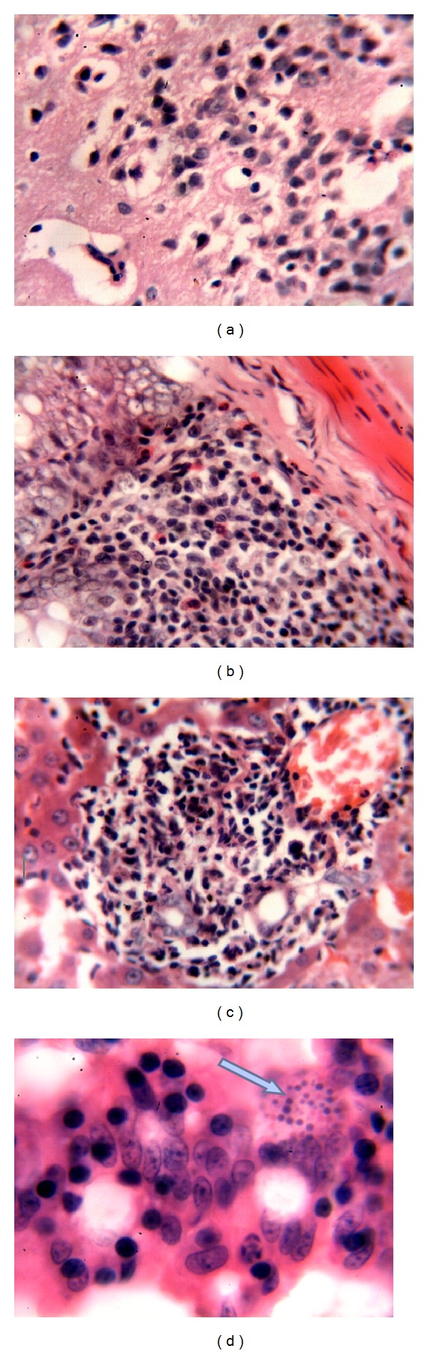
This study was performed to evaluate pathology of experimental Encephalitozoon cuniculi (Iraqi isolate) infection in normal and immunosuppressed mice. Pathological changes were not seen in negative control mice while secondary bacterial infections were noted in the lungs, kidneys, and heart of mice given dexamethasone. Typical E. cuniculi infection lesions were found in brain, livers, lungs, and kidneys of mice given 10(7) E. cuniculi spores/mouse orally. These lesions were in the form of nonsuppurative meningoencephalitis with vasculitis in brain, interstitial inflammation with infiltration of both lymphocytes and plasma cells in lung tissue, and nonsuppurative interstitial (focal and diffuse) nephritis, presence of vacuole containing mature and immature spores in enterocytes within the tips of villi, and lymphoiod hyperplasia of the white pulp and vasculitis of the intratrabecular vessels. Mice that were given 10(7) E. cuniculi spores/mouse orally showed lesions similar to those observed in the previous group (vasculitis and granulomas) but the lesions were more severe and widespread. In conclusion, this is the first report of experimental E. cuniculi infection induced by E. cuniculi isolated from a naturally infected rabbit in Iraq and that infection became more severe and widespread upon the administration of dexaethasone.

 求助内容:
求助内容: 应助结果提醒方式:


