{"title":"41例寄生源可触及浅表结节的细胞学研究。","authors":"Prashant Goyal, Shelly Sehgal, Soumyesh Ghosh, Deepti Mittal, Awanindra Kumar, Sompal Singh","doi":"10.1155/2014/373472","DOIUrl":null,"url":null,"abstract":"<p><p>Background. Few parasitic infestations present as only superficial palpable subcutaneous or intramuscular nodule. The current study highlights the role of FNAC in the diagnosis of superficial palpable parasitic lesions. Methods. This was a retrospective study in which we reviewed the FNAC record of all patients over a period of two years from September 2011 to August 2013. During this period, FNA was performed on 5954 cases which presented as superficial palpable lump at various sites of body. There were 41 cases diagnosed as parasitic lesion or suspicious of parasitic lesion on cytology which were included in the study. Results. In the present study, most of the patients were children and young adults. The lesions were located over trunk in 18 (43.9%) cases, extremities in 12 (29.3%) cases, and head and neck region in 11 (26.8%) cases. Out of 41 cases, 27 (65.8%) cases were confirmed on cytology and/or histopathology as parasitic lesions, including 21 (51.2%) cases of cysticercosis, 5 (12.2%) cases of filariasis, and one (2.4%) case of hydatid cyst. Cytological findings of remaining cases were suggestive of parasitic lesion. Conclusion. Careful assessment of cytological material is helpful to detect parasite or inflammatory response to parasite even in asymptomatic patients. </p>","PeriodicalId":89212,"journal":{"name":"Pathology research international","volume":"2014 ","pages":"373472"},"PeriodicalIF":0.0000,"publicationDate":"2014-01-01","publicationTypes":"Journal Article","fieldsOfStudy":null,"isOpenAccess":false,"openAccessPdf":"https://sci-hub-pdf.com/10.1155/2014/373472","citationCount":"18","resultStr":"{\"title\":\"A cytological study of palpable superficial nodules of parasitic origin: a study of 41 cases.\",\"authors\":\"Prashant Goyal, Shelly Sehgal, Soumyesh Ghosh, Deepti Mittal, Awanindra Kumar, Sompal Singh\",\"doi\":\"10.1155/2014/373472\",\"DOIUrl\":null,\"url\":null,\"abstract\":\"<p><p>Background. Few parasitic infestations present as only superficial palpable subcutaneous or intramuscular nodule. The current study highlights the role of FNAC in the diagnosis of superficial palpable parasitic lesions. Methods. This was a retrospective study in which we reviewed the FNAC record of all patients over a period of two years from September 2011 to August 2013. During this period, FNA was performed on 5954 cases which presented as superficial palpable lump at various sites of body. There were 41 cases diagnosed as parasitic lesion or suspicious of parasitic lesion on cytology which were included in the study. Results. In the present study, most of the patients were children and young adults. The lesions were located over trunk in 18 (43.9%) cases, extremities in 12 (29.3%) cases, and head and neck region in 11 (26.8%) cases. Out of 41 cases, 27 (65.8%) cases were confirmed on cytology and/or histopathology as parasitic lesions, including 21 (51.2%) cases of cysticercosis, 5 (12.2%) cases of filariasis, and one (2.4%) case of hydatid cyst. Cytological findings of remaining cases were suggestive of parasitic lesion. Conclusion. Careful assessment of cytological material is helpful to detect parasite or inflammatory response to parasite even in asymptomatic patients. </p>\",\"PeriodicalId\":89212,\"journal\":{\"name\":\"Pathology research international\",\"volume\":\"2014 \",\"pages\":\"373472\"},\"PeriodicalIF\":0.0000,\"publicationDate\":\"2014-01-01\",\"publicationTypes\":\"Journal Article\",\"fieldsOfStudy\":null,\"isOpenAccess\":false,\"openAccessPdf\":\"https://sci-hub-pdf.com/10.1155/2014/373472\",\"citationCount\":\"18\",\"resultStr\":null,\"platform\":\"Semanticscholar\",\"paperid\":null,\"PeriodicalName\":\"Pathology research international\",\"FirstCategoryId\":\"1085\",\"ListUrlMain\":\"https://doi.org/10.1155/2014/373472\",\"RegionNum\":0,\"RegionCategory\":null,\"ArticlePicture\":[],\"TitleCN\":null,\"AbstractTextCN\":null,\"PMCID\":null,\"EPubDate\":\"2014/3/17 0:00:00\",\"PubModel\":\"Epub\",\"JCR\":\"\",\"JCRName\":\"\",\"Score\":null,\"Total\":0}","platform":"Semanticscholar","paperid":null,"PeriodicalName":"Pathology research international","FirstCategoryId":"1085","ListUrlMain":"https://doi.org/10.1155/2014/373472","RegionNum":0,"RegionCategory":null,"ArticlePicture":[],"TitleCN":null,"AbstractTextCN":null,"PMCID":null,"EPubDate":"2014/3/17 0:00:00","PubModel":"Epub","JCR":"","JCRName":"","Score":null,"Total":0}
A cytological study of palpable superficial nodules of parasitic origin: a study of 41 cases.
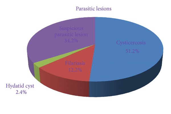
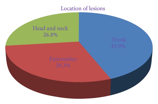
Background. Few parasitic infestations present as only superficial palpable subcutaneous or intramuscular nodule. The current study highlights the role of FNAC in the diagnosis of superficial palpable parasitic lesions. Methods. This was a retrospective study in which we reviewed the FNAC record of all patients over a period of two years from September 2011 to August 2013. During this period, FNA was performed on 5954 cases which presented as superficial palpable lump at various sites of body. There were 41 cases diagnosed as parasitic lesion or suspicious of parasitic lesion on cytology which were included in the study. Results. In the present study, most of the patients were children and young adults. The lesions were located over trunk in 18 (43.9%) cases, extremities in 12 (29.3%) cases, and head and neck region in 11 (26.8%) cases. Out of 41 cases, 27 (65.8%) cases were confirmed on cytology and/or histopathology as parasitic lesions, including 21 (51.2%) cases of cysticercosis, 5 (12.2%) cases of filariasis, and one (2.4%) case of hydatid cyst. Cytological findings of remaining cases were suggestive of parasitic lesion. Conclusion. Careful assessment of cytological material is helpful to detect parasite or inflammatory response to parasite even in asymptomatic patients.


 求助内容:
求助内容: 应助结果提醒方式:


