Santam Chakraborty, Firuza D Patel, Vijay M Patil, Arun S Oinam, Suresh C Sharma
{"title":"宫颈癌高剂量率近距离放疗期间器官剂量干扰变化的幅度和意义:一项基于CT的计划研究。","authors":"Santam Chakraborty, Firuza D Patel, Vijay M Patil, Arun S Oinam, Suresh C Sharma","doi":"10.1155/2014/687365","DOIUrl":null,"url":null,"abstract":"<p><p>Background. Quantifying the interfraction dose variations in the organs at risk (OAR) in HDR intracavitary brachytherapy (HDR ICBT). Methods. Rectum and bladder were contoured in 44 patients of cervical carcinoma on CT after each fraction of HDR ICBT (9 Gy/2 fractions). Interfraction dose variations (VARact) were calculated. Rigid image registration of consecutive fraction images allowed quantification of the hypothetical variation in dose (VARhypo) arising exclusively due to changes in applicator placement and geometry. VARhypo was regressed against the VARact to find out to what extent the applicator variation could explain the VARact in the OAR. The rest of the variation was assumed to be due to organ deformation. Results. The VARact in the dose to 2 cc of bladder and rectum were 1.46 and 1.16 Gy, respectively. Increased dose was seen in 16 and 23 patients in the subsequent fraction for bladder and rectum, respectively. Doses to OAR would have exceeded constraints in 16% patients if second fraction was not imaged. VARhypo explained 19% and 47% of the VARact observed for the bladder and rectum respectively. Conclusions. Significant interfraction variations in OAR doses can occur in HDR ICBT. Organ deformations are mostly responsible for this variation. </p>","PeriodicalId":89399,"journal":{"name":"ISRN oncology","volume":"2014 ","pages":"687365"},"PeriodicalIF":0.0000,"publicationDate":"2014-02-03","publicationTypes":"Journal Article","fieldsOfStudy":null,"isOpenAccess":false,"openAccessPdf":"https://sci-hub-pdf.com/10.1155/2014/687365","citationCount":"17","resultStr":"{\"title\":\"Magnitude and Implications of Interfraction Variations in Organ Doses during High Dose Rate Brachytherapy of Cervix Cancer: A CT Based Planning Study.\",\"authors\":\"Santam Chakraborty, Firuza D Patel, Vijay M Patil, Arun S Oinam, Suresh C Sharma\",\"doi\":\"10.1155/2014/687365\",\"DOIUrl\":null,\"url\":null,\"abstract\":\"<p><p>Background. Quantifying the interfraction dose variations in the organs at risk (OAR) in HDR intracavitary brachytherapy (HDR ICBT). Methods. Rectum and bladder were contoured in 44 patients of cervical carcinoma on CT after each fraction of HDR ICBT (9 Gy/2 fractions). Interfraction dose variations (VARact) were calculated. Rigid image registration of consecutive fraction images allowed quantification of the hypothetical variation in dose (VARhypo) arising exclusively due to changes in applicator placement and geometry. VARhypo was regressed against the VARact to find out to what extent the applicator variation could explain the VARact in the OAR. The rest of the variation was assumed to be due to organ deformation. Results. The VARact in the dose to 2 cc of bladder and rectum were 1.46 and 1.16 Gy, respectively. Increased dose was seen in 16 and 23 patients in the subsequent fraction for bladder and rectum, respectively. Doses to OAR would have exceeded constraints in 16% patients if second fraction was not imaged. VARhypo explained 19% and 47% of the VARact observed for the bladder and rectum respectively. Conclusions. Significant interfraction variations in OAR doses can occur in HDR ICBT. Organ deformations are mostly responsible for this variation. </p>\",\"PeriodicalId\":89399,\"journal\":{\"name\":\"ISRN oncology\",\"volume\":\"2014 \",\"pages\":\"687365\"},\"PeriodicalIF\":0.0000,\"publicationDate\":\"2014-02-03\",\"publicationTypes\":\"Journal Article\",\"fieldsOfStudy\":null,\"isOpenAccess\":false,\"openAccessPdf\":\"https://sci-hub-pdf.com/10.1155/2014/687365\",\"citationCount\":\"17\",\"resultStr\":null,\"platform\":\"Semanticscholar\",\"paperid\":null,\"PeriodicalName\":\"ISRN oncology\",\"FirstCategoryId\":\"1085\",\"ListUrlMain\":\"https://doi.org/10.1155/2014/687365\",\"RegionNum\":0,\"RegionCategory\":null,\"ArticlePicture\":[],\"TitleCN\":null,\"AbstractTextCN\":null,\"PMCID\":null,\"EPubDate\":\"2014/1/1 0:00:00\",\"PubModel\":\"eCollection\",\"JCR\":\"\",\"JCRName\":\"\",\"Score\":null,\"Total\":0}","platform":"Semanticscholar","paperid":null,"PeriodicalName":"ISRN oncology","FirstCategoryId":"1085","ListUrlMain":"https://doi.org/10.1155/2014/687365","RegionNum":0,"RegionCategory":null,"ArticlePicture":[],"TitleCN":null,"AbstractTextCN":null,"PMCID":null,"EPubDate":"2014/1/1 0:00:00","PubModel":"eCollection","JCR":"","JCRName":"","Score":null,"Total":0}
Magnitude and Implications of Interfraction Variations in Organ Doses during High Dose Rate Brachytherapy of Cervix Cancer: A CT Based Planning Study.
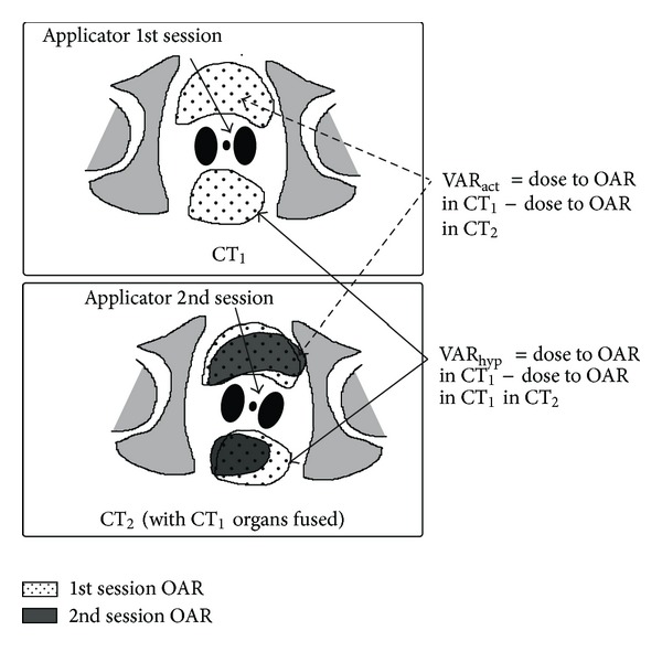
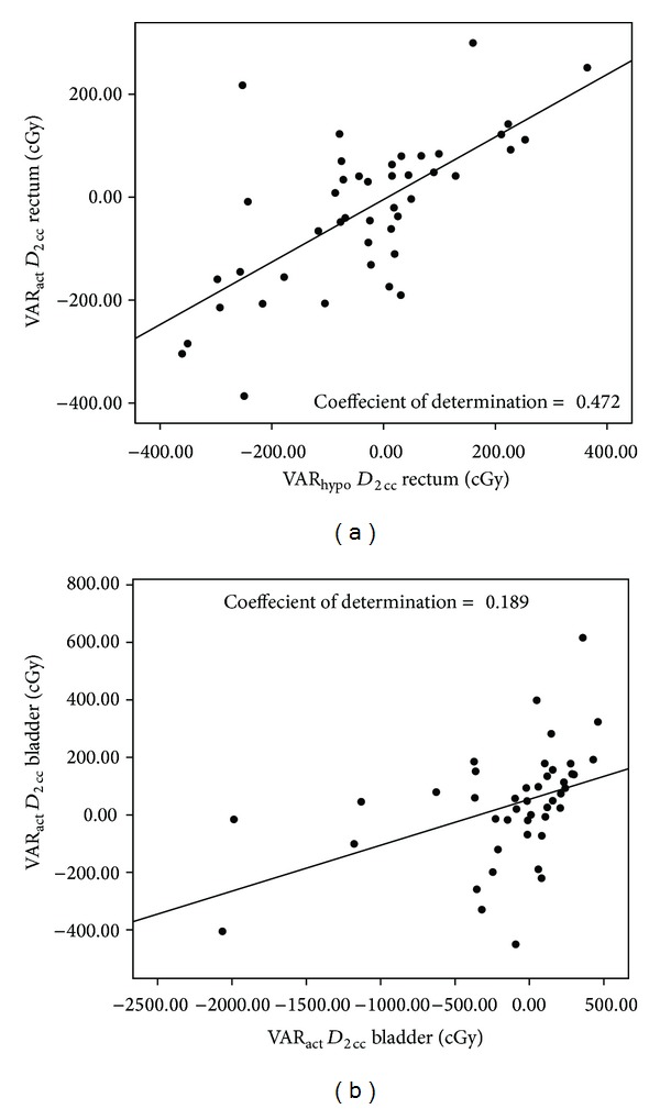
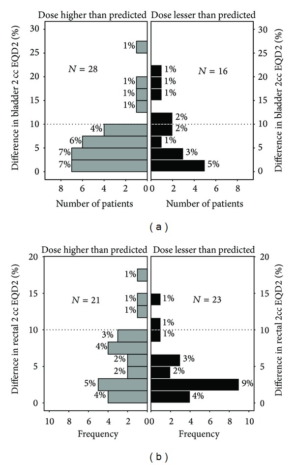
Background. Quantifying the interfraction dose variations in the organs at risk (OAR) in HDR intracavitary brachytherapy (HDR ICBT). Methods. Rectum and bladder were contoured in 44 patients of cervical carcinoma on CT after each fraction of HDR ICBT (9 Gy/2 fractions). Interfraction dose variations (VARact) were calculated. Rigid image registration of consecutive fraction images allowed quantification of the hypothetical variation in dose (VARhypo) arising exclusively due to changes in applicator placement and geometry. VARhypo was regressed against the VARact to find out to what extent the applicator variation could explain the VARact in the OAR. The rest of the variation was assumed to be due to organ deformation. Results. The VARact in the dose to 2 cc of bladder and rectum were 1.46 and 1.16 Gy, respectively. Increased dose was seen in 16 and 23 patients in the subsequent fraction for bladder and rectum, respectively. Doses to OAR would have exceeded constraints in 16% patients if second fraction was not imaged. VARhypo explained 19% and 47% of the VARact observed for the bladder and rectum respectively. Conclusions. Significant interfraction variations in OAR doses can occur in HDR ICBT. Organ deformations are mostly responsible for this variation.

 求助内容:
求助内容: 应助结果提醒方式:


