María Del Carmen Baez, Mariana Tarán, Candelaria Llorens, Ariel Balceda, María de La Paz Scribano, Patricia Pons, Mónica Moya
{"title":"大鼠主动脉平滑肌细胞线粒体形态功能的改变。","authors":"María Del Carmen Baez, Mariana Tarán, Candelaria Llorens, Ariel Balceda, María de La Paz Scribano, Patricia Pons, Mónica Moya","doi":"10.1155/2014/739526","DOIUrl":null,"url":null,"abstract":"<p><p>In an experimental model of atherogenesis induced by hyperfibrinogenemia (HF), the pharmacological response of vitamin E was studied in order to assess its antioxidant effect on the mitochondrial morphofunctional alterations in aortic smooth muscle cells. Three groups of male rats were used: (Ctr) control, (AI) atherogenesis induced for 120 days, and (AIE) atherogenesis induced for 120 days and treated with vitamin E. HF was induced by adrenalin injection (0.1 mg/day/rat) for 120 days. AIE group was treated with the administration of 3.42 mg/day/rat of vitamin E for 105 days after the first induction. Mitochondria morphology was analyzed by electronic microscopy (EM) and mitochondrial complexes (MC) by spectrophotometry. In group AI the total and mean number of mitochondria reduced significantly, the intermembranous matrix increased, and swelling was observed with respect to Ctr and AIE (P < 0.01). These damages were related to a significant decrease in the activity of citrate synthase and complexes I, II, III, and IV in group AI in comparison to Ctr (P < 0.001). Similar behavior was presented by group AI compared to AIE (P < 0.001). These results show that vitamin E produces a significative regression of inflammatory and oxidative stress process and it resolved the morphofunctional mitochondrial alterations in this experimental model of atherogenic disease. </p>","PeriodicalId":73519,"journal":{"name":"ISRN cardiology","volume":"2014 ","pages":"739526"},"PeriodicalIF":0.0000,"publicationDate":"2014-02-06","publicationTypes":"Journal Article","fieldsOfStudy":null,"isOpenAccess":false,"openAccessPdf":"https://sci-hub-pdf.com/10.1155/2014/739526","citationCount":"1","resultStr":"{\"title\":\"Mitochondrial morphofunctional alterations in smooth muscle cells of aorta in rats.\",\"authors\":\"María Del Carmen Baez, Mariana Tarán, Candelaria Llorens, Ariel Balceda, María de La Paz Scribano, Patricia Pons, Mónica Moya\",\"doi\":\"10.1155/2014/739526\",\"DOIUrl\":null,\"url\":null,\"abstract\":\"<p><p>In an experimental model of atherogenesis induced by hyperfibrinogenemia (HF), the pharmacological response of vitamin E was studied in order to assess its antioxidant effect on the mitochondrial morphofunctional alterations in aortic smooth muscle cells. Three groups of male rats were used: (Ctr) control, (AI) atherogenesis induced for 120 days, and (AIE) atherogenesis induced for 120 days and treated with vitamin E. HF was induced by adrenalin injection (0.1 mg/day/rat) for 120 days. AIE group was treated with the administration of 3.42 mg/day/rat of vitamin E for 105 days after the first induction. Mitochondria morphology was analyzed by electronic microscopy (EM) and mitochondrial complexes (MC) by spectrophotometry. In group AI the total and mean number of mitochondria reduced significantly, the intermembranous matrix increased, and swelling was observed with respect to Ctr and AIE (P < 0.01). These damages were related to a significant decrease in the activity of citrate synthase and complexes I, II, III, and IV in group AI in comparison to Ctr (P < 0.001). Similar behavior was presented by group AI compared to AIE (P < 0.001). These results show that vitamin E produces a significative regression of inflammatory and oxidative stress process and it resolved the morphofunctional mitochondrial alterations in this experimental model of atherogenic disease. </p>\",\"PeriodicalId\":73519,\"journal\":{\"name\":\"ISRN cardiology\",\"volume\":\"2014 \",\"pages\":\"739526\"},\"PeriodicalIF\":0.0000,\"publicationDate\":\"2014-02-06\",\"publicationTypes\":\"Journal Article\",\"fieldsOfStudy\":null,\"isOpenAccess\":false,\"openAccessPdf\":\"https://sci-hub-pdf.com/10.1155/2014/739526\",\"citationCount\":\"1\",\"resultStr\":null,\"platform\":\"Semanticscholar\",\"paperid\":null,\"PeriodicalName\":\"ISRN cardiology\",\"FirstCategoryId\":\"1085\",\"ListUrlMain\":\"https://doi.org/10.1155/2014/739526\",\"RegionNum\":0,\"RegionCategory\":null,\"ArticlePicture\":[],\"TitleCN\":null,\"AbstractTextCN\":null,\"PMCID\":null,\"EPubDate\":\"2014/1/1 0:00:00\",\"PubModel\":\"eCollection\",\"JCR\":\"\",\"JCRName\":\"\",\"Score\":null,\"Total\":0}","platform":"Semanticscholar","paperid":null,"PeriodicalName":"ISRN cardiology","FirstCategoryId":"1085","ListUrlMain":"https://doi.org/10.1155/2014/739526","RegionNum":0,"RegionCategory":null,"ArticlePicture":[],"TitleCN":null,"AbstractTextCN":null,"PMCID":null,"EPubDate":"2014/1/1 0:00:00","PubModel":"eCollection","JCR":"","JCRName":"","Score":null,"Total":0}
Mitochondrial morphofunctional alterations in smooth muscle cells of aorta in rats.
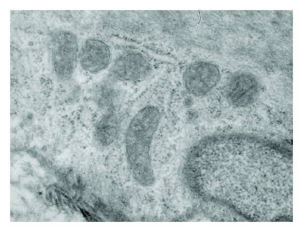
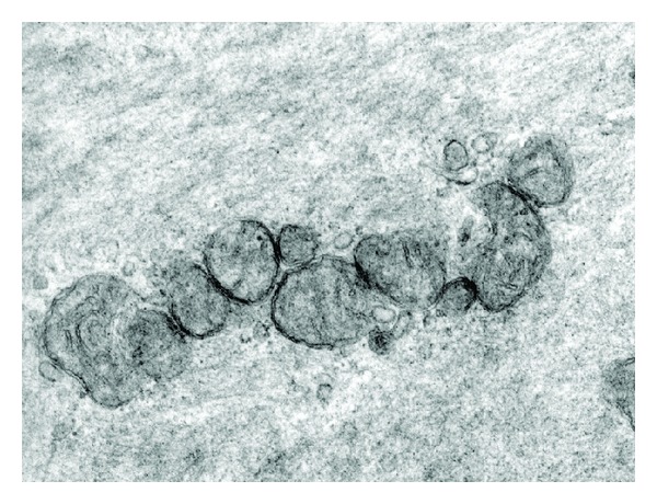
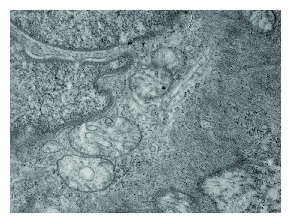
In an experimental model of atherogenesis induced by hyperfibrinogenemia (HF), the pharmacological response of vitamin E was studied in order to assess its antioxidant effect on the mitochondrial morphofunctional alterations in aortic smooth muscle cells. Three groups of male rats were used: (Ctr) control, (AI) atherogenesis induced for 120 days, and (AIE) atherogenesis induced for 120 days and treated with vitamin E. HF was induced by adrenalin injection (0.1 mg/day/rat) for 120 days. AIE group was treated with the administration of 3.42 mg/day/rat of vitamin E for 105 days after the first induction. Mitochondria morphology was analyzed by electronic microscopy (EM) and mitochondrial complexes (MC) by spectrophotometry. In group AI the total and mean number of mitochondria reduced significantly, the intermembranous matrix increased, and swelling was observed with respect to Ctr and AIE (P < 0.01). These damages were related to a significant decrease in the activity of citrate synthase and complexes I, II, III, and IV in group AI in comparison to Ctr (P < 0.001). Similar behavior was presented by group AI compared to AIE (P < 0.001). These results show that vitamin E produces a significative regression of inflammatory and oxidative stress process and it resolved the morphofunctional mitochondrial alterations in this experimental model of atherogenic disease.

 求助内容:
求助内容: 应助结果提醒方式:


