{"title":"术中前段光学相干断层扫描在Descemet剥离自动内皮角膜移植术中的疗效。","authors":"Akio Miyakoshi, Hironori Ozaki, Mitsuya Otsuka, Atsushi Hayashi","doi":"10.1155/2014/562062","DOIUrl":null,"url":null,"abstract":"<p><p>Purpose. To examine the graft-host interface during Descemet's stripping automated endothelial keratoplasty (DSAEK) surgery with optical coherence tomography (OCT). Design. Prospective, interventional case series. Patients and Methods. Eight patients who underwent a DSAEK were included. A handheld OCT was used intraoperatively to examine the presence of interface fluid between the host cornea and the graft. Results. In 3 patients, no interface fluid was detected between the host cornea and the graft after the graft was attached by air injection. In 4 patients, interface fluid was detected after the graft was attached by air injection. The remaining interface fluid was drained through corneal stab incisions. One patient required a second surgery because the first surgery failed due to persistence of the interface fluid. All patients showed a complete attachment of the graft at one month after the DSAEK surgery. Conclusion. A handheld OCT is useful to detect the interface fluid between the host cornea and the graft during a DSAEK. </p>","PeriodicalId":90193,"journal":{"name":"ISRN ophthalmology","volume":"2014 ","pages":"562062"},"PeriodicalIF":0.0000,"publicationDate":"2014-02-02","publicationTypes":"Journal Article","fieldsOfStudy":null,"isOpenAccess":false,"openAccessPdf":"https://sci-hub-pdf.com/10.1155/2014/562062","citationCount":"17","resultStr":"{\"title\":\"Efficacy of Intraoperative Anterior Segment Optical Coherence Tomography during Descemet's Stripping Automated Endothelial Keratoplasty.\",\"authors\":\"Akio Miyakoshi, Hironori Ozaki, Mitsuya Otsuka, Atsushi Hayashi\",\"doi\":\"10.1155/2014/562062\",\"DOIUrl\":null,\"url\":null,\"abstract\":\"<p><p>Purpose. To examine the graft-host interface during Descemet's stripping automated endothelial keratoplasty (DSAEK) surgery with optical coherence tomography (OCT). Design. Prospective, interventional case series. Patients and Methods. Eight patients who underwent a DSAEK were included. A handheld OCT was used intraoperatively to examine the presence of interface fluid between the host cornea and the graft. Results. In 3 patients, no interface fluid was detected between the host cornea and the graft after the graft was attached by air injection. In 4 patients, interface fluid was detected after the graft was attached by air injection. The remaining interface fluid was drained through corneal stab incisions. One patient required a second surgery because the first surgery failed due to persistence of the interface fluid. All patients showed a complete attachment of the graft at one month after the DSAEK surgery. Conclusion. A handheld OCT is useful to detect the interface fluid between the host cornea and the graft during a DSAEK. </p>\",\"PeriodicalId\":90193,\"journal\":{\"name\":\"ISRN ophthalmology\",\"volume\":\"2014 \",\"pages\":\"562062\"},\"PeriodicalIF\":0.0000,\"publicationDate\":\"2014-02-02\",\"publicationTypes\":\"Journal Article\",\"fieldsOfStudy\":null,\"isOpenAccess\":false,\"openAccessPdf\":\"https://sci-hub-pdf.com/10.1155/2014/562062\",\"citationCount\":\"17\",\"resultStr\":null,\"platform\":\"Semanticscholar\",\"paperid\":null,\"PeriodicalName\":\"ISRN ophthalmology\",\"FirstCategoryId\":\"1085\",\"ListUrlMain\":\"https://doi.org/10.1155/2014/562062\",\"RegionNum\":0,\"RegionCategory\":null,\"ArticlePicture\":[],\"TitleCN\":null,\"AbstractTextCN\":null,\"PMCID\":null,\"EPubDate\":\"2014/1/1 0:00:00\",\"PubModel\":\"eCollection\",\"JCR\":\"\",\"JCRName\":\"\",\"Score\":null,\"Total\":0}","platform":"Semanticscholar","paperid":null,"PeriodicalName":"ISRN ophthalmology","FirstCategoryId":"1085","ListUrlMain":"https://doi.org/10.1155/2014/562062","RegionNum":0,"RegionCategory":null,"ArticlePicture":[],"TitleCN":null,"AbstractTextCN":null,"PMCID":null,"EPubDate":"2014/1/1 0:00:00","PubModel":"eCollection","JCR":"","JCRName":"","Score":null,"Total":0}
Efficacy of Intraoperative Anterior Segment Optical Coherence Tomography during Descemet's Stripping Automated Endothelial Keratoplasty.
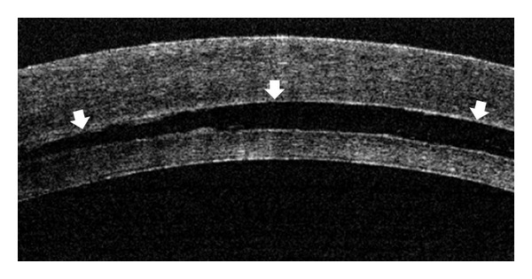
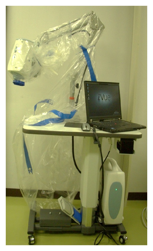
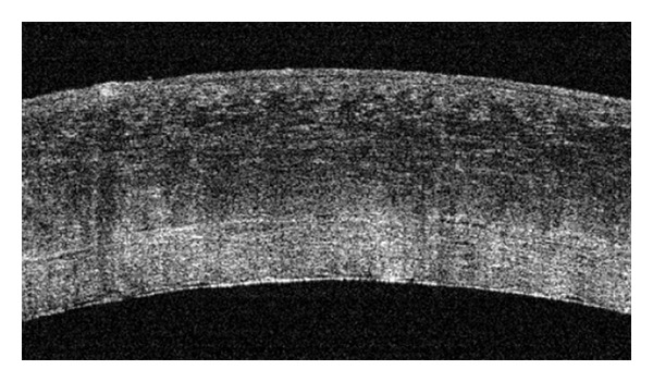
Purpose. To examine the graft-host interface during Descemet's stripping automated endothelial keratoplasty (DSAEK) surgery with optical coherence tomography (OCT). Design. Prospective, interventional case series. Patients and Methods. Eight patients who underwent a DSAEK were included. A handheld OCT was used intraoperatively to examine the presence of interface fluid between the host cornea and the graft. Results. In 3 patients, no interface fluid was detected between the host cornea and the graft after the graft was attached by air injection. In 4 patients, interface fluid was detected after the graft was attached by air injection. The remaining interface fluid was drained through corneal stab incisions. One patient required a second surgery because the first surgery failed due to persistence of the interface fluid. All patients showed a complete attachment of the graft at one month after the DSAEK surgery. Conclusion. A handheld OCT is useful to detect the interface fluid between the host cornea and the graft during a DSAEK.

 求助内容:
求助内容: 应助结果提醒方式:


