Sergei I Agoulnik, Satoshi Kawano, Noel Taylor, Judith Oestreicher, Junji Matsui, Jesse Chow, Yoshiya Oda, Yasuhiro Funahashi
{"title":"甲磺酸埃立布林在体外引起周细胞特异性基因表达改变,缩短周细胞驱动的毛细血管网络。","authors":"Sergei I Agoulnik, Satoshi Kawano, Noel Taylor, Judith Oestreicher, Junji Matsui, Jesse Chow, Yoshiya Oda, Yasuhiro Funahashi","doi":"10.1186/2045-824X-6-3","DOIUrl":null,"url":null,"abstract":"<p><strong>Background: </strong>Eribulin mesylate is a synthetic macrocyclic ketone analog of the marine sponge natural product halichondrin B. Eribulin is a tubulin-binding drug and approved in many countries worldwide for treatment of certain patients with advanced breast cancer. Here we investigated antiproliferative and antiangiogenic effects of eribulin on vascular cells, human umbilical vein endothelial cells (HUVECs) and human brain vascular pericytes (HBVPs), in vitro in comparison with another tubulin-binding drug, paclitaxel.</p><p><strong>Methods: </strong>HUVECs and HBVPs were treated with either eribulin or paclitaxel and their antiproliferative effects were evaluated. Global gene expression profiling changes caused by drug treatments were studied using Affymetrix microarray platform and custom TaqMan Low Density Cards. To examine effects of the drugs on pericyte-driven in vitro angiogenesis, we compared lengths of capillary networks in co-cultures of HUVECs with HBVPs.</p><p><strong>Results: </strong>Both eribulin and paclitaxel showed potent activities in in vitro proliferation of HUVECs and HBVPs, with the half-maximal inhibitory concentrations (IC50) in low- to sub-nmol/L concentrations. When gene expression changes were assessed in HUVECs, the majority of affected genes overlapped for both treatments (59%), while in HBVPs, altered gene signatures were drug-dependent and the overlap was limited to just 12%. In HBVPs, eribulin selectively affected 11 pathways (p < 0.01) such as Cell Cycle Control of Chromosomal Replication. In contrast, paclitaxel was tended to regulate 27 pathways such as PI3K/AKT. Only 5 pathways were commonly affected by both treatments. In in vitro pericyte-driven angiogenesis model, paclitaxel showed limited activity while eribulin shortened the formed capillary networks of HUVECs driven by HBVPs at low nmol/L concentrations starting at day 3 after treatments.</p><p><strong>Conclusions: </strong>Our findings suggest that pericytes, but not endothelial cells, responded differently, to two mechanistically-distinct tubulin-binding drugs, eribulin and paclitaxel. While eribulin and paclitaxel induced similar changes in gene expression in endothelial cells, in pericytes their altered gene expression was unique and drug-specific. In the functional endothelial-pericyte co-culture assay, eribulin, but not paclitaxel showed strong efficacy not only as a cytotoxic drug but also as a potent antivascular agent that affected pericyte-driven in vitro angiogenesis.</p>","PeriodicalId":23948,"journal":{"name":"Vascular Cell","volume":"6 1","pages":"3"},"PeriodicalIF":0.0000,"publicationDate":"2014-03-01","publicationTypes":"Journal Article","fieldsOfStudy":null,"isOpenAccess":false,"openAccessPdf":"https://sci-hub-pdf.com/10.1186/2045-824X-6-3","citationCount":"35","resultStr":"{\"title\":\"Eribulin mesylate exerts specific gene expression changes in pericytes and shortens pericyte-driven capillary network in vitro.\",\"authors\":\"Sergei I Agoulnik, Satoshi Kawano, Noel Taylor, Judith Oestreicher, Junji Matsui, Jesse Chow, Yoshiya Oda, Yasuhiro Funahashi\",\"doi\":\"10.1186/2045-824X-6-3\",\"DOIUrl\":null,\"url\":null,\"abstract\":\"<p><strong>Background: </strong>Eribulin mesylate is a synthetic macrocyclic ketone analog of the marine sponge natural product halichondrin B. Eribulin is a tubulin-binding drug and approved in many countries worldwide for treatment of certain patients with advanced breast cancer. Here we investigated antiproliferative and antiangiogenic effects of eribulin on vascular cells, human umbilical vein endothelial cells (HUVECs) and human brain vascular pericytes (HBVPs), in vitro in comparison with another tubulin-binding drug, paclitaxel.</p><p><strong>Methods: </strong>HUVECs and HBVPs were treated with either eribulin or paclitaxel and their antiproliferative effects were evaluated. Global gene expression profiling changes caused by drug treatments were studied using Affymetrix microarray platform and custom TaqMan Low Density Cards. To examine effects of the drugs on pericyte-driven in vitro angiogenesis, we compared lengths of capillary networks in co-cultures of HUVECs with HBVPs.</p><p><strong>Results: </strong>Both eribulin and paclitaxel showed potent activities in in vitro proliferation of HUVECs and HBVPs, with the half-maximal inhibitory concentrations (IC50) in low- to sub-nmol/L concentrations. When gene expression changes were assessed in HUVECs, the majority of affected genes overlapped for both treatments (59%), while in HBVPs, altered gene signatures were drug-dependent and the overlap was limited to just 12%. In HBVPs, eribulin selectively affected 11 pathways (p < 0.01) such as Cell Cycle Control of Chromosomal Replication. In contrast, paclitaxel was tended to regulate 27 pathways such as PI3K/AKT. Only 5 pathways were commonly affected by both treatments. In in vitro pericyte-driven angiogenesis model, paclitaxel showed limited activity while eribulin shortened the formed capillary networks of HUVECs driven by HBVPs at low nmol/L concentrations starting at day 3 after treatments.</p><p><strong>Conclusions: </strong>Our findings suggest that pericytes, but not endothelial cells, responded differently, to two mechanistically-distinct tubulin-binding drugs, eribulin and paclitaxel. While eribulin and paclitaxel induced similar changes in gene expression in endothelial cells, in pericytes their altered gene expression was unique and drug-specific. In the functional endothelial-pericyte co-culture assay, eribulin, but not paclitaxel showed strong efficacy not only as a cytotoxic drug but also as a potent antivascular agent that affected pericyte-driven in vitro angiogenesis.</p>\",\"PeriodicalId\":23948,\"journal\":{\"name\":\"Vascular Cell\",\"volume\":\"6 1\",\"pages\":\"3\"},\"PeriodicalIF\":0.0000,\"publicationDate\":\"2014-03-01\",\"publicationTypes\":\"Journal Article\",\"fieldsOfStudy\":null,\"isOpenAccess\":false,\"openAccessPdf\":\"https://sci-hub-pdf.com/10.1186/2045-824X-6-3\",\"citationCount\":\"35\",\"resultStr\":null,\"platform\":\"Semanticscholar\",\"paperid\":null,\"PeriodicalName\":\"Vascular Cell\",\"FirstCategoryId\":\"1085\",\"ListUrlMain\":\"https://doi.org/10.1186/2045-824X-6-3\",\"RegionNum\":0,\"RegionCategory\":null,\"ArticlePicture\":[],\"TitleCN\":null,\"AbstractTextCN\":null,\"PMCID\":null,\"EPubDate\":\"\",\"PubModel\":\"\",\"JCR\":\"Q4\",\"JCRName\":\"Neuroscience\",\"Score\":null,\"Total\":0}","platform":"Semanticscholar","paperid":null,"PeriodicalName":"Vascular Cell","FirstCategoryId":"1085","ListUrlMain":"https://doi.org/10.1186/2045-824X-6-3","RegionNum":0,"RegionCategory":null,"ArticlePicture":[],"TitleCN":null,"AbstractTextCN":null,"PMCID":null,"EPubDate":"","PubModel":"","JCR":"Q4","JCRName":"Neuroscience","Score":null,"Total":0}
Eribulin mesylate exerts specific gene expression changes in pericytes and shortens pericyte-driven capillary network in vitro.
Background: Eribulin mesylate is a synthetic macrocyclic ketone analog of the marine sponge natural product halichondrin B. Eribulin is a tubulin-binding drug and approved in many countries worldwide for treatment of certain patients with advanced breast cancer. Here we investigated antiproliferative and antiangiogenic effects of eribulin on vascular cells, human umbilical vein endothelial cells (HUVECs) and human brain vascular pericytes (HBVPs), in vitro in comparison with another tubulin-binding drug, paclitaxel.
Methods: HUVECs and HBVPs were treated with either eribulin or paclitaxel and their antiproliferative effects were evaluated. Global gene expression profiling changes caused by drug treatments were studied using Affymetrix microarray platform and custom TaqMan Low Density Cards. To examine effects of the drugs on pericyte-driven in vitro angiogenesis, we compared lengths of capillary networks in co-cultures of HUVECs with HBVPs.
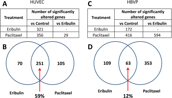
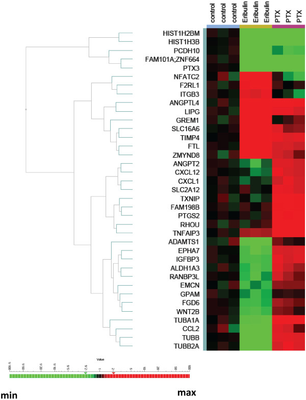
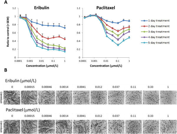
Results: Both eribulin and paclitaxel showed potent activities in in vitro proliferation of HUVECs and HBVPs, with the half-maximal inhibitory concentrations (IC50) in low- to sub-nmol/L concentrations. When gene expression changes were assessed in HUVECs, the majority of affected genes overlapped for both treatments (59%), while in HBVPs, altered gene signatures were drug-dependent and the overlap was limited to just 12%. In HBVPs, eribulin selectively affected 11 pathways (p < 0.01) such as Cell Cycle Control of Chromosomal Replication. In contrast, paclitaxel was tended to regulate 27 pathways such as PI3K/AKT. Only 5 pathways were commonly affected by both treatments. In in vitro pericyte-driven angiogenesis model, paclitaxel showed limited activity while eribulin shortened the formed capillary networks of HUVECs driven by HBVPs at low nmol/L concentrations starting at day 3 after treatments.
Conclusions: Our findings suggest that pericytes, but not endothelial cells, responded differently, to two mechanistically-distinct tubulin-binding drugs, eribulin and paclitaxel. While eribulin and paclitaxel induced similar changes in gene expression in endothelial cells, in pericytes their altered gene expression was unique and drug-specific. In the functional endothelial-pericyte co-culture assay, eribulin, but not paclitaxel showed strong efficacy not only as a cytotoxic drug but also as a potent antivascular agent that affected pericyte-driven in vitro angiogenesis.

 求助内容:
求助内容: 应助结果提醒方式:


