Peter C Marks, Marilena Preda, Terry Henderson, Lucy Liaw, Volkhard Lindner, Robert E Friesel, Ilka M Pinz
{"title":"小鼠后肢缺血模型磁共振血管成像血管树和侧支血管体积的交互式三维分析。","authors":"Peter C Marks, Marilena Preda, Terry Henderson, Lucy Liaw, Volkhard Lindner, Robert E Friesel, Ilka M Pinz","doi":"10.2174/1874347101307010019","DOIUrl":null,"url":null,"abstract":"<p><p>The quantitative analysis of blood vessel volumes from magnetic resonance angiograms (MRA) or μCT images is difficult and time-consuming. This fact, when combined with a study that involves multiple scans of multiple subjects, can represent a significant portion of research time. In order to enhance analysis options and to provide an automated and fast analysis method, we developed a software plugin for the ImageJ and Fiji image processing frameworks that enables the quick and reproducible volume quantification of blood vessel segments. The novel plugin named Volume Calculator (VolCal), accepts any binary (thresholded) image and produces a three-dimensional schematic representation of the vasculature that can be directly manipulated by the investigator. Using MRAs of the mouse hindlimb ischemia model, we demonstrate quick and reproducible blood vessel volume calculations with 95 - 98% accuracy. In clinical settings this software may enhance image interpretation and the speed of data analysis and thus enhance intervention decisions for example in peripheral vascular disease or aneurysms. In summary, we provide a novel, fast and interactive quantification of blood vessel volumes for single blood vessels or sets of vessel segments with particular focus on collateral formation after an ischemic insult.</p>","PeriodicalId":90366,"journal":{"name":"The open medical imaging journal","volume":"7 ","pages":"19-27"},"PeriodicalIF":0.0000,"publicationDate":"2013-10-31","publicationTypes":"Journal Article","fieldsOfStudy":null,"isOpenAccess":false,"openAccessPdf":"https://www.ncbi.nlm.nih.gov/pmc/articles/PMC3929959/pdf/","citationCount":"11","resultStr":"{\"title\":\"Interactive 3D Analysis of Blood Vessel Trees and Collateral Vessel Volumes in Magnetic Resonance Angiograms in the Mouse Ischemic Hindlimb Model.\",\"authors\":\"Peter C Marks, Marilena Preda, Terry Henderson, Lucy Liaw, Volkhard Lindner, Robert E Friesel, Ilka M Pinz\",\"doi\":\"10.2174/1874347101307010019\",\"DOIUrl\":null,\"url\":null,\"abstract\":\"<p><p>The quantitative analysis of blood vessel volumes from magnetic resonance angiograms (MRA) or μCT images is difficult and time-consuming. This fact, when combined with a study that involves multiple scans of multiple subjects, can represent a significant portion of research time. In order to enhance analysis options and to provide an automated and fast analysis method, we developed a software plugin for the ImageJ and Fiji image processing frameworks that enables the quick and reproducible volume quantification of blood vessel segments. The novel plugin named Volume Calculator (VolCal), accepts any binary (thresholded) image and produces a three-dimensional schematic representation of the vasculature that can be directly manipulated by the investigator. Using MRAs of the mouse hindlimb ischemia model, we demonstrate quick and reproducible blood vessel volume calculations with 95 - 98% accuracy. In clinical settings this software may enhance image interpretation and the speed of data analysis and thus enhance intervention decisions for example in peripheral vascular disease or aneurysms. In summary, we provide a novel, fast and interactive quantification of blood vessel volumes for single blood vessels or sets of vessel segments with particular focus on collateral formation after an ischemic insult.</p>\",\"PeriodicalId\":90366,\"journal\":{\"name\":\"The open medical imaging journal\",\"volume\":\"7 \",\"pages\":\"19-27\"},\"PeriodicalIF\":0.0000,\"publicationDate\":\"2013-10-31\",\"publicationTypes\":\"Journal Article\",\"fieldsOfStudy\":null,\"isOpenAccess\":false,\"openAccessPdf\":\"https://www.ncbi.nlm.nih.gov/pmc/articles/PMC3929959/pdf/\",\"citationCount\":\"11\",\"resultStr\":null,\"platform\":\"Semanticscholar\",\"paperid\":null,\"PeriodicalName\":\"The open medical imaging journal\",\"FirstCategoryId\":\"1085\",\"ListUrlMain\":\"https://doi.org/10.2174/1874347101307010019\",\"RegionNum\":0,\"RegionCategory\":null,\"ArticlePicture\":[],\"TitleCN\":null,\"AbstractTextCN\":null,\"PMCID\":null,\"EPubDate\":\"\",\"PubModel\":\"\",\"JCR\":\"\",\"JCRName\":\"\",\"Score\":null,\"Total\":0}","platform":"Semanticscholar","paperid":null,"PeriodicalName":"The open medical imaging journal","FirstCategoryId":"1085","ListUrlMain":"https://doi.org/10.2174/1874347101307010019","RegionNum":0,"RegionCategory":null,"ArticlePicture":[],"TitleCN":null,"AbstractTextCN":null,"PMCID":null,"EPubDate":"","PubModel":"","JCR":"","JCRName":"","Score":null,"Total":0}
Interactive 3D Analysis of Blood Vessel Trees and Collateral Vessel Volumes in Magnetic Resonance Angiograms in the Mouse Ischemic Hindlimb Model.
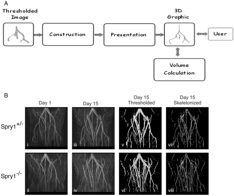
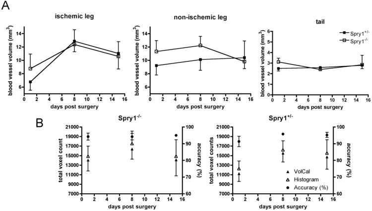
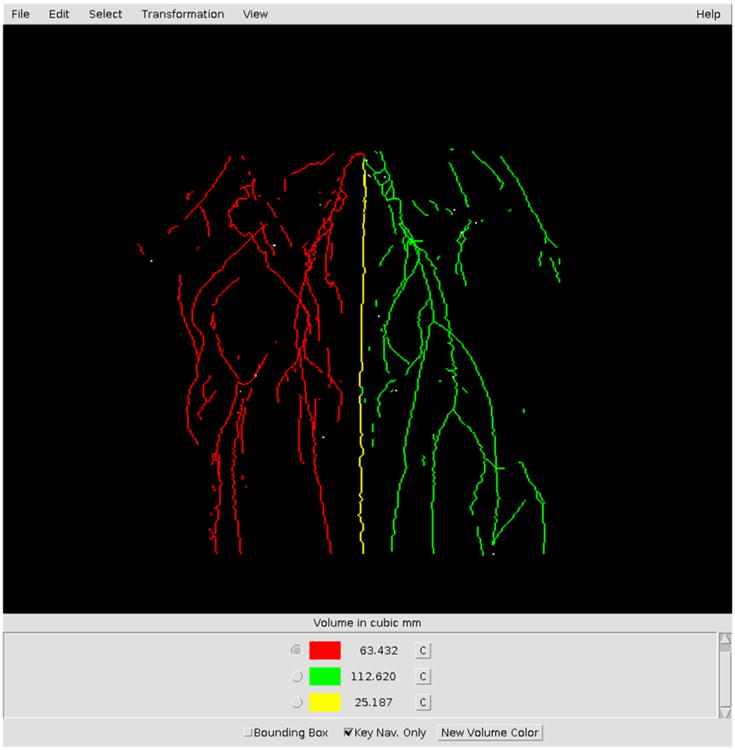
The quantitative analysis of blood vessel volumes from magnetic resonance angiograms (MRA) or μCT images is difficult and time-consuming. This fact, when combined with a study that involves multiple scans of multiple subjects, can represent a significant portion of research time. In order to enhance analysis options and to provide an automated and fast analysis method, we developed a software plugin for the ImageJ and Fiji image processing frameworks that enables the quick and reproducible volume quantification of blood vessel segments. The novel plugin named Volume Calculator (VolCal), accepts any binary (thresholded) image and produces a three-dimensional schematic representation of the vasculature that can be directly manipulated by the investigator. Using MRAs of the mouse hindlimb ischemia model, we demonstrate quick and reproducible blood vessel volume calculations with 95 - 98% accuracy. In clinical settings this software may enhance image interpretation and the speed of data analysis and thus enhance intervention decisions for example in peripheral vascular disease or aneurysms. In summary, we provide a novel, fast and interactive quantification of blood vessel volumes for single blood vessels or sets of vessel segments with particular focus on collateral formation after an ischemic insult.

 求助内容:
求助内容: 应助结果提醒方式:


