Flore De Bats, Benjamin Wolff, Martine Mauget-Faÿsse, Isabelle Meunier, Philippe Denis, Laurent Kodjikian
{"title":"网状假性早衰与早发性早衰的关系。","authors":"Flore De Bats, Benjamin Wolff, Martine Mauget-Faÿsse, Isabelle Meunier, Philippe Denis, Laurent Kodjikian","doi":"10.1155/2013/273085","DOIUrl":null,"url":null,"abstract":"<p><p>Purpose. To report an association between reticular pseudodrusen, located above the retinal pigment epithelium (RPE), and Early Onset Drusen (EOD) as described using Spectral-Domain Optical Coherence Tomography (SD-OCT). Methods. Eight patients (16 eyes) with EOD were examined. EOD were classified into three entities called Large Colloid Drusen (LCD), Malattia Leventinese (ML), and Cuticular Drusen (CD). Best-corrected visual acuity, fundus examination, color fundus photographs, fundus autofluorescence (FAF), fluorescein angiography (FA), indocyanine green angiography (ICGA), and SD-OCT were performed in all study patients. Results. Four patients had LCD, 2 had ML, and 2 had CD. Reticular pseudodrusen were observed with SD-OCT in all study patients; all these patients had hyperreflective lesions above and below the RPE. Conclusion. Early Onset Drusen appear to be associated with reticular pseudodrusen. SD-OCT is helpful in distinguishing the location of the deposits that are above and below the RPE in EOD. Further studies are needed to understand the role of reticular pseudodrusen in the pathophysiology of EOD. </p>","PeriodicalId":90193,"journal":{"name":"ISRN ophthalmology","volume":"2013 ","pages":"273085"},"PeriodicalIF":0.0000,"publicationDate":"2013-05-16","publicationTypes":"Journal Article","fieldsOfStudy":null,"isOpenAccess":false,"openAccessPdf":"https://sci-hub-pdf.com/10.1155/2013/273085","citationCount":"20","resultStr":"{\"title\":\"Association of reticular pseudodrusen and early onset drusen.\",\"authors\":\"Flore De Bats, Benjamin Wolff, Martine Mauget-Faÿsse, Isabelle Meunier, Philippe Denis, Laurent Kodjikian\",\"doi\":\"10.1155/2013/273085\",\"DOIUrl\":null,\"url\":null,\"abstract\":\"<p><p>Purpose. To report an association between reticular pseudodrusen, located above the retinal pigment epithelium (RPE), and Early Onset Drusen (EOD) as described using Spectral-Domain Optical Coherence Tomography (SD-OCT). Methods. Eight patients (16 eyes) with EOD were examined. EOD were classified into three entities called Large Colloid Drusen (LCD), Malattia Leventinese (ML), and Cuticular Drusen (CD). Best-corrected visual acuity, fundus examination, color fundus photographs, fundus autofluorescence (FAF), fluorescein angiography (FA), indocyanine green angiography (ICGA), and SD-OCT were performed in all study patients. Results. Four patients had LCD, 2 had ML, and 2 had CD. Reticular pseudodrusen were observed with SD-OCT in all study patients; all these patients had hyperreflective lesions above and below the RPE. Conclusion. Early Onset Drusen appear to be associated with reticular pseudodrusen. SD-OCT is helpful in distinguishing the location of the deposits that are above and below the RPE in EOD. Further studies are needed to understand the role of reticular pseudodrusen in the pathophysiology of EOD. </p>\",\"PeriodicalId\":90193,\"journal\":{\"name\":\"ISRN ophthalmology\",\"volume\":\"2013 \",\"pages\":\"273085\"},\"PeriodicalIF\":0.0000,\"publicationDate\":\"2013-05-16\",\"publicationTypes\":\"Journal Article\",\"fieldsOfStudy\":null,\"isOpenAccess\":false,\"openAccessPdf\":\"https://sci-hub-pdf.com/10.1155/2013/273085\",\"citationCount\":\"20\",\"resultStr\":null,\"platform\":\"Semanticscholar\",\"paperid\":null,\"PeriodicalName\":\"ISRN ophthalmology\",\"FirstCategoryId\":\"1085\",\"ListUrlMain\":\"https://doi.org/10.1155/2013/273085\",\"RegionNum\":0,\"RegionCategory\":null,\"ArticlePicture\":[],\"TitleCN\":null,\"AbstractTextCN\":null,\"PMCID\":null,\"EPubDate\":\"2013/1/1 0:00:00\",\"PubModel\":\"eCollection\",\"JCR\":\"\",\"JCRName\":\"\",\"Score\":null,\"Total\":0}","platform":"Semanticscholar","paperid":null,"PeriodicalName":"ISRN ophthalmology","FirstCategoryId":"1085","ListUrlMain":"https://doi.org/10.1155/2013/273085","RegionNum":0,"RegionCategory":null,"ArticlePicture":[],"TitleCN":null,"AbstractTextCN":null,"PMCID":null,"EPubDate":"2013/1/1 0:00:00","PubModel":"eCollection","JCR":"","JCRName":"","Score":null,"Total":0}
Association of reticular pseudodrusen and early onset drusen.
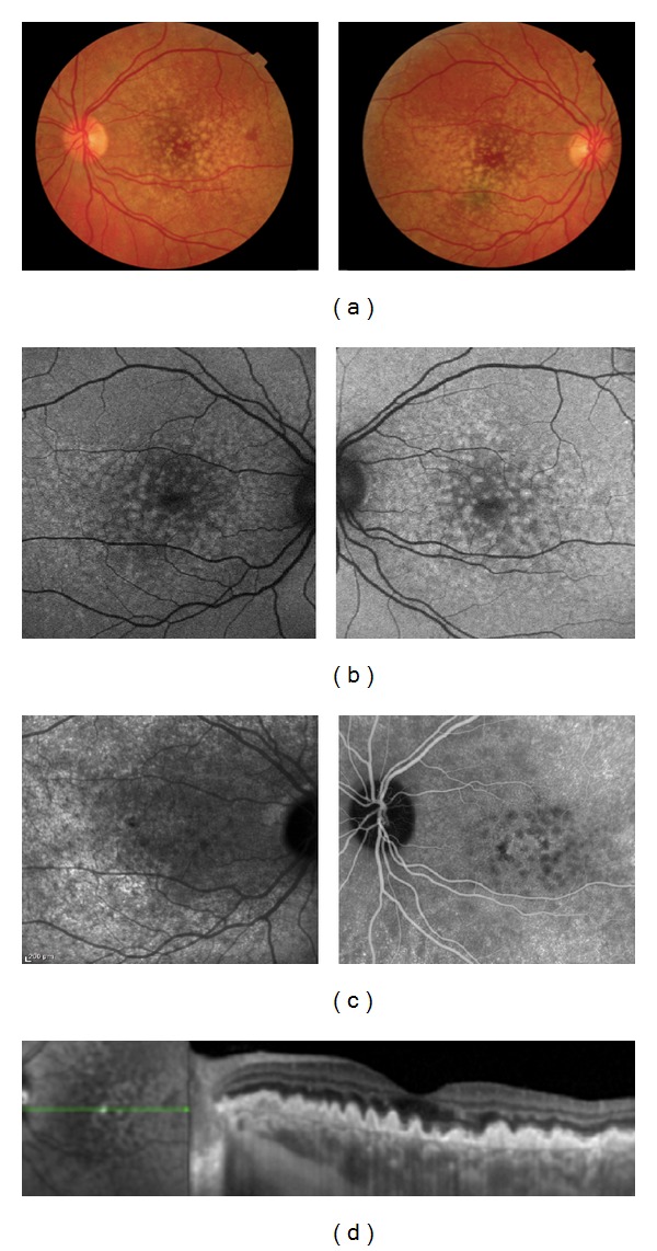
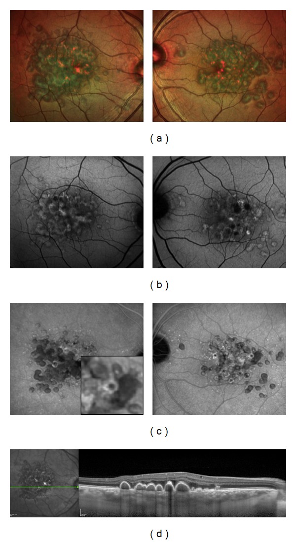
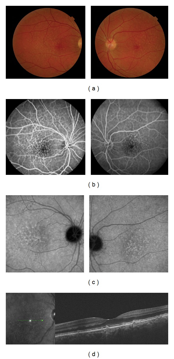
Purpose. To report an association between reticular pseudodrusen, located above the retinal pigment epithelium (RPE), and Early Onset Drusen (EOD) as described using Spectral-Domain Optical Coherence Tomography (SD-OCT). Methods. Eight patients (16 eyes) with EOD were examined. EOD were classified into three entities called Large Colloid Drusen (LCD), Malattia Leventinese (ML), and Cuticular Drusen (CD). Best-corrected visual acuity, fundus examination, color fundus photographs, fundus autofluorescence (FAF), fluorescein angiography (FA), indocyanine green angiography (ICGA), and SD-OCT were performed in all study patients. Results. Four patients had LCD, 2 had ML, and 2 had CD. Reticular pseudodrusen were observed with SD-OCT in all study patients; all these patients had hyperreflective lesions above and below the RPE. Conclusion. Early Onset Drusen appear to be associated with reticular pseudodrusen. SD-OCT is helpful in distinguishing the location of the deposits that are above and below the RPE in EOD. Further studies are needed to understand the role of reticular pseudodrusen in the pathophysiology of EOD.

 求助内容:
求助内容: 应助结果提醒方式:


