Maria L Salvetat, Marco Zeppieri, Flavia Miani, Paolo Brusini
{"title":"角膜移植术前后角膜表面波前分析:Descemet剥离自动内皮角膜移植术与穿透性角膜移植术。","authors":"Maria L Salvetat, Marco Zeppieri, Flavia Miani, Paolo Brusini","doi":"10.1155/2013/210565","DOIUrl":null,"url":null,"abstract":"<p><p>Purpose. To compare the higher-order aberrations (HOAs) due to the anterior and posterior corneal surfaces in patients that underwent either Descemet-stripping-automated-endothelial-keratoplasty (DSAEK) or penetrating keratoplasty (PK) for endothelial dysfunction and age-matched controls. Methods. This retrospective, observational, case series included 28 patients after PK, 30 patients after DSAEK, and 30 healthy controls. A Scheimpflug imaging system was used to assess the HOAs due to the anterior and posterior corneal surfaces at 4 mm and 6 mm optical zones. Total, 3rd and 4th order HOAs were considered. Intra- and intergroup differences were assessed using the Friedman and the Kruskal-Wallis tests, respectively; paired comparisons were performed using Duncan's multiple range test. Results. Total, 3rd and 4th order HOAs due to both corneal surfaces at 4 mm and 6 mm optical zones were significantly higher in the PK group, intermediate in the DSAEK group, and lower in controls (P < 0.05). The most important HOAs components in both PK and DSAEK groups were trefoil and coma from the anterior corneal surface (P < 0.05) and trefoil from the posterior corneal surface (P < 0.05). Conclusions. The optical quality of both corneal surfaces appeared significantly higher after DSAEK than after PK, which can increase the postoperative patient's quality of vision and satisfaction. </p>","PeriodicalId":90193,"journal":{"name":"ISRN ophthalmology","volume":"2013 ","pages":"210565"},"PeriodicalIF":0.0000,"publicationDate":"2013-09-12","publicationTypes":"Journal Article","fieldsOfStudy":null,"isOpenAccess":false,"openAccessPdf":"https://sci-hub-pdf.com/10.1155/2013/210565","citationCount":"1","resultStr":"{\"title\":\"Postkeratoplasty Anterior and Posterior Corneal Surface Wavefront Analysis: Descemet's Stripping Automated Endothelial Keratoplasty versus Penetrating Keratoplasty.\",\"authors\":\"Maria L Salvetat, Marco Zeppieri, Flavia Miani, Paolo Brusini\",\"doi\":\"10.1155/2013/210565\",\"DOIUrl\":null,\"url\":null,\"abstract\":\"<p><p>Purpose. To compare the higher-order aberrations (HOAs) due to the anterior and posterior corneal surfaces in patients that underwent either Descemet-stripping-automated-endothelial-keratoplasty (DSAEK) or penetrating keratoplasty (PK) for endothelial dysfunction and age-matched controls. Methods. This retrospective, observational, case series included 28 patients after PK, 30 patients after DSAEK, and 30 healthy controls. A Scheimpflug imaging system was used to assess the HOAs due to the anterior and posterior corneal surfaces at 4 mm and 6 mm optical zones. Total, 3rd and 4th order HOAs were considered. Intra- and intergroup differences were assessed using the Friedman and the Kruskal-Wallis tests, respectively; paired comparisons were performed using Duncan's multiple range test. Results. Total, 3rd and 4th order HOAs due to both corneal surfaces at 4 mm and 6 mm optical zones were significantly higher in the PK group, intermediate in the DSAEK group, and lower in controls (P < 0.05). The most important HOAs components in both PK and DSAEK groups were trefoil and coma from the anterior corneal surface (P < 0.05) and trefoil from the posterior corneal surface (P < 0.05). Conclusions. The optical quality of both corneal surfaces appeared significantly higher after DSAEK than after PK, which can increase the postoperative patient's quality of vision and satisfaction. </p>\",\"PeriodicalId\":90193,\"journal\":{\"name\":\"ISRN ophthalmology\",\"volume\":\"2013 \",\"pages\":\"210565\"},\"PeriodicalIF\":0.0000,\"publicationDate\":\"2013-09-12\",\"publicationTypes\":\"Journal Article\",\"fieldsOfStudy\":null,\"isOpenAccess\":false,\"openAccessPdf\":\"https://sci-hub-pdf.com/10.1155/2013/210565\",\"citationCount\":\"1\",\"resultStr\":null,\"platform\":\"Semanticscholar\",\"paperid\":null,\"PeriodicalName\":\"ISRN ophthalmology\",\"FirstCategoryId\":\"1085\",\"ListUrlMain\":\"https://doi.org/10.1155/2013/210565\",\"RegionNum\":0,\"RegionCategory\":null,\"ArticlePicture\":[],\"TitleCN\":null,\"AbstractTextCN\":null,\"PMCID\":null,\"EPubDate\":\"2013/1/1 0:00:00\",\"PubModel\":\"eCollection\",\"JCR\":\"\",\"JCRName\":\"\",\"Score\":null,\"Total\":0}","platform":"Semanticscholar","paperid":null,"PeriodicalName":"ISRN ophthalmology","FirstCategoryId":"1085","ListUrlMain":"https://doi.org/10.1155/2013/210565","RegionNum":0,"RegionCategory":null,"ArticlePicture":[],"TitleCN":null,"AbstractTextCN":null,"PMCID":null,"EPubDate":"2013/1/1 0:00:00","PubModel":"eCollection","JCR":"","JCRName":"","Score":null,"Total":0}
Postkeratoplasty Anterior and Posterior Corneal Surface Wavefront Analysis: Descemet's Stripping Automated Endothelial Keratoplasty versus Penetrating Keratoplasty.
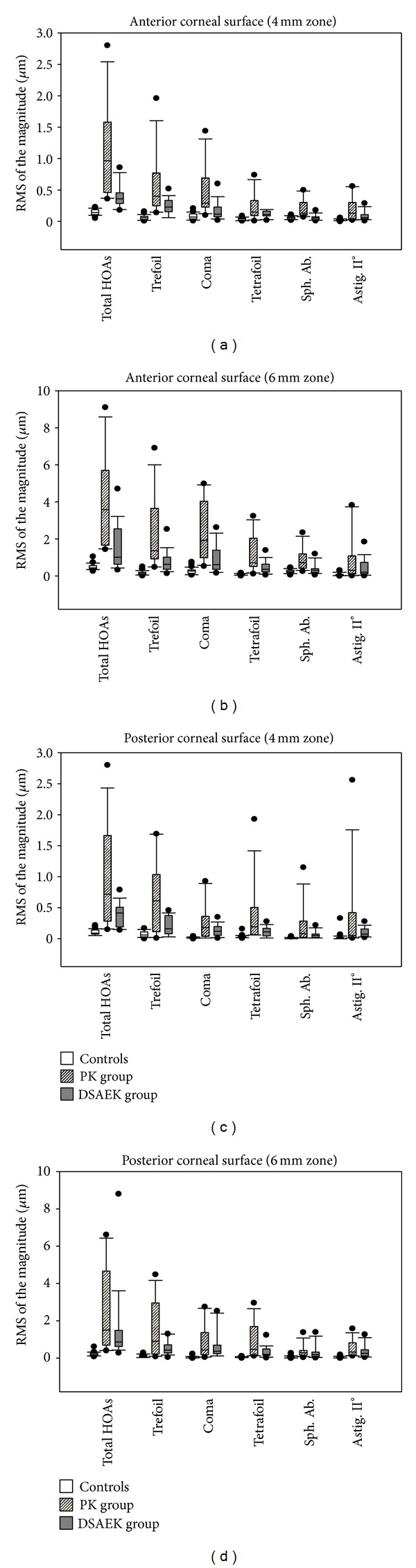
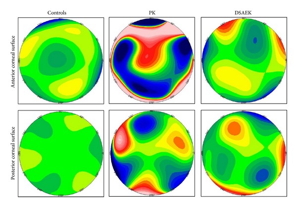
Purpose. To compare the higher-order aberrations (HOAs) due to the anterior and posterior corneal surfaces in patients that underwent either Descemet-stripping-automated-endothelial-keratoplasty (DSAEK) or penetrating keratoplasty (PK) for endothelial dysfunction and age-matched controls. Methods. This retrospective, observational, case series included 28 patients after PK, 30 patients after DSAEK, and 30 healthy controls. A Scheimpflug imaging system was used to assess the HOAs due to the anterior and posterior corneal surfaces at 4 mm and 6 mm optical zones. Total, 3rd and 4th order HOAs were considered. Intra- and intergroup differences were assessed using the Friedman and the Kruskal-Wallis tests, respectively; paired comparisons were performed using Duncan's multiple range test. Results. Total, 3rd and 4th order HOAs due to both corneal surfaces at 4 mm and 6 mm optical zones were significantly higher in the PK group, intermediate in the DSAEK group, and lower in controls (P < 0.05). The most important HOAs components in both PK and DSAEK groups were trefoil and coma from the anterior corneal surface (P < 0.05) and trefoil from the posterior corneal surface (P < 0.05). Conclusions. The optical quality of both corneal surfaces appeared significantly higher after DSAEK than after PK, which can increase the postoperative patient's quality of vision and satisfaction.

 求助内容:
求助内容: 应助结果提醒方式:


