Kayoung Yi, Mircea Mujat, Wei Sun, B Hyle Park, Johannes F de Boer, Teresa C Chen
{"title":"评估青光眼患者的乳头周围视网膜厚度图:一个新概念。","authors":"Kayoung Yi, Mircea Mujat, Wei Sun, B Hyle Park, Johannes F de Boer, Teresa C Chen","doi":"10.5402/2011/146813","DOIUrl":null,"url":null,"abstract":"<p><p>Purpose. To show how peripapillary spectral domain optical coherence tomography (SDOCT) retinal thickness (RT) maps can complement retinal nerve fiber layer (RNFL) thickness maps in the evaluation of glaucoma patients. Methods. After a complete eye exam with standard fundus photography and visual field testing, normal and glaucomatous eyes were imaged with an experimental SDOCT system. From SDOCT images, RNFL thickness and RT maps were constructed and then correlated with disc photography and visual field testing. Results. Two normal eyes of 2 patients and 5 eyes of 4 glaucoma patients were imaged. Although both RNFL and RT maps correlated well with visual field defects, glaucomatous arcuate defects were sometimes more easily identified in the RT maps. Conclusions. To our knowledge, this is the first paper to show that peripapillary SDOCT RT maps may provide important supplemental information to RNFL thickness maps in the evaluation of glaucoma patients. </p>","PeriodicalId":90193,"journal":{"name":"ISRN ophthalmology","volume":"2011 ","pages":"146813"},"PeriodicalIF":0.0000,"publicationDate":"2011-09-07","publicationTypes":"Journal Article","fieldsOfStudy":null,"isOpenAccess":false,"openAccessPdf":"https://www.ncbi.nlm.nih.gov/pmc/articles/PMC3912594/pdf/","citationCount":"8","resultStr":"{\"title\":\"Peripapillary retinal thickness maps in the evaluation of glaucoma patients: a novel concept.\",\"authors\":\"Kayoung Yi, Mircea Mujat, Wei Sun, B Hyle Park, Johannes F de Boer, Teresa C Chen\",\"doi\":\"10.5402/2011/146813\",\"DOIUrl\":null,\"url\":null,\"abstract\":\"<p><p>Purpose. To show how peripapillary spectral domain optical coherence tomography (SDOCT) retinal thickness (RT) maps can complement retinal nerve fiber layer (RNFL) thickness maps in the evaluation of glaucoma patients. Methods. After a complete eye exam with standard fundus photography and visual field testing, normal and glaucomatous eyes were imaged with an experimental SDOCT system. From SDOCT images, RNFL thickness and RT maps were constructed and then correlated with disc photography and visual field testing. Results. Two normal eyes of 2 patients and 5 eyes of 4 glaucoma patients were imaged. Although both RNFL and RT maps correlated well with visual field defects, glaucomatous arcuate defects were sometimes more easily identified in the RT maps. Conclusions. To our knowledge, this is the first paper to show that peripapillary SDOCT RT maps may provide important supplemental information to RNFL thickness maps in the evaluation of glaucoma patients. </p>\",\"PeriodicalId\":90193,\"journal\":{\"name\":\"ISRN ophthalmology\",\"volume\":\"2011 \",\"pages\":\"146813\"},\"PeriodicalIF\":0.0000,\"publicationDate\":\"2011-09-07\",\"publicationTypes\":\"Journal Article\",\"fieldsOfStudy\":null,\"isOpenAccess\":false,\"openAccessPdf\":\"https://www.ncbi.nlm.nih.gov/pmc/articles/PMC3912594/pdf/\",\"citationCount\":\"8\",\"resultStr\":null,\"platform\":\"Semanticscholar\",\"paperid\":null,\"PeriodicalName\":\"ISRN ophthalmology\",\"FirstCategoryId\":\"1085\",\"ListUrlMain\":\"https://doi.org/10.5402/2011/146813\",\"RegionNum\":0,\"RegionCategory\":null,\"ArticlePicture\":[],\"TitleCN\":null,\"AbstractTextCN\":null,\"PMCID\":null,\"EPubDate\":\"2011/1/1 0:00:00\",\"PubModel\":\"eCollection\",\"JCR\":\"\",\"JCRName\":\"\",\"Score\":null,\"Total\":0}","platform":"Semanticscholar","paperid":null,"PeriodicalName":"ISRN ophthalmology","FirstCategoryId":"1085","ListUrlMain":"https://doi.org/10.5402/2011/146813","RegionNum":0,"RegionCategory":null,"ArticlePicture":[],"TitleCN":null,"AbstractTextCN":null,"PMCID":null,"EPubDate":"2011/1/1 0:00:00","PubModel":"eCollection","JCR":"","JCRName":"","Score":null,"Total":0}
Peripapillary retinal thickness maps in the evaluation of glaucoma patients: a novel concept.
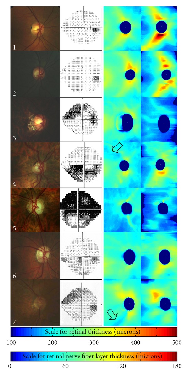
Purpose. To show how peripapillary spectral domain optical coherence tomography (SDOCT) retinal thickness (RT) maps can complement retinal nerve fiber layer (RNFL) thickness maps in the evaluation of glaucoma patients. Methods. After a complete eye exam with standard fundus photography and visual field testing, normal and glaucomatous eyes were imaged with an experimental SDOCT system. From SDOCT images, RNFL thickness and RT maps were constructed and then correlated with disc photography and visual field testing. Results. Two normal eyes of 2 patients and 5 eyes of 4 glaucoma patients were imaged. Although both RNFL and RT maps correlated well with visual field defects, glaucomatous arcuate defects were sometimes more easily identified in the RT maps. Conclusions. To our knowledge, this is the first paper to show that peripapillary SDOCT RT maps may provide important supplemental information to RNFL thickness maps in the evaluation of glaucoma patients.

 求助内容:
求助内容: 应助结果提醒方式:


